
 E-mail Alert
 RSS
RSS
| Citation: |

|
Automatic 3D vertebrae CT image active contour segmentation method based on weighted random forest
-
Abstract
In order to solve the problems of sensitive initial contours and inaccurate segmentation caused by active contour segmentation of CT images, this paper proposes an automatic 3D vertebral CT active contour segmentation method combined weighted random forest called "WRF-AC". This method proposes a weighted random forest algorithm and an active contour energy function that includes edge energy. First, the weighted random forest is trained by extracting 3D Haar-like feature values of the vertebra CT, and the 'vertebra center' obtained is used as the initial contour of the segmentation. Then, the segmentation of the vertebra CT image is completed by solving the active contour energy function minimum containing the edge energy. The experimental results show that this method can segment the spine CT images more accurately and quickly on the same datasets to extract the vertebrae.-
Keywords:
- 3D segmentation /
- CT images /
- weighted random forest /
- active contour
-

-
References
[1] Roth H R, Farag A, Lu L, et al. Deep convolutional networks for pancreas segmentation in CT imaging[J]. Proceedings of SPIE, 2015, 9413: 94131G. [2] Glocker B, Feulner J, Criminisi A, et al. Automatic localization and identification of vertebrae in arbitrary field-of-view CT scans[M]//Ayache N, Delingette H, Golland P, et al. Medical Image Computing and Computer-Assisted Intervention. Berlin Heidelberg: Springer, 2012: 590–598. [3] Kang Y, Engelke K, Kalender W A. A new accurate and precise 3-D segmentation method for skeletal structures in volumetric CT data[J]. IEEE Transactions Medical Imaging, 2003, 22(5): 586–598. doi: 10.1109/TMI.2003.812265 [4] Lim P H, Bagci U, Bai L. Introducing Willmore flow into level set segmentation of spinal vertebrae[J]. IEEE Transactions on Biomedical Engineering, 2013, 60(1): 115–122. [5] Huang J Y, Jian F Z, Wu H, et al. An improved level set method for vertebra CT image segmentation[J]. Biomedical Engineering Online, 2013, 12: 48. doi: 10.1186/1475-925X-12-48 [6] 唐利明, 田学全, 黄大荣, 等.结合FCMS与变分水平集的图像分割模型[J].自动化学报, 2014, 40(6): 1233–1248. Tang L M, Tian X Q, Huang D R, et al. Image segmentation model combined with FCMS and variational level set[J]. Acta Automatica Sinica, 2014, 40(6): 1233–1248. [7] Criminisi A, Shotton J, Robertson D, et al. Regression forests for efficient anatomy detection and localization in CT studies[M]//Menze B, Langs G, Tu Z W, et al. Medical Computer Vision. Recognition Techniques and Applications in Medical Imaging. Berlin Heidelberg: Springer, 2010: 106–117. [8] Criminisi A, Robertson D, Konukoglu E, et al. Regression forests for efficient anatomy detection and localization in computed tomography scans[J]. Medical Image Analysis, 2013, 17(8): 1293–1303. doi: 10.1016/j.media.2013.01.001 [9] Cuingnet R, Prevost R, Lesage D, et al. Automatic detection and segmentation of kidneys in 3D CT images using random forests[M]//Ayache N, Delingette H, Golland P, et al. Medical Image Computing and Computer-Assisted Intervention. Berlin Heidelberg: Springer, 2012: 66–74. [10] Klinder T, Ostermann J, Ehm M, et al. Automated model-based vertebra detection, identification, and segmentation in CT images[J]. Medical Image Analysis, 2009, 13(3): 471–482. [11] Huang S H, Chu Y H, Lai S H, et al. Learning-based vertebra detection and iterative normalized-cut segmentation for spinal MRI[J]. IEEE Transactions on Medical Imaging, 2009, 28(10): 1595–1605. doi: 10.1109/TMI.2009.2023362 [12] Roberts M G, Cootes T F, Pacheco E, et al. Segmentation of lumbar vertebrae using part-based graphs and active appearance models[M]//Yang G Z, Hawkes D, Rueckert D, et al. Medical Image Computing and Computer-Assisted Intervention. Berlin Heidelberg: Springer, 2009: 1017–1024. [13] 陈侃, 李彬, 田联房.基于模糊速度函数的活动轮廓模型的肺结节分割[J].自动化学报, 2013, 39(8): 1257–1264. Chen K, Li B, Tian L F. A segmentation algorithm of pulmonary nodules using active contour model based on fuzzy speed function[J]. Acta Automatica Sinica, 2013, 39(8): 1257–1264. [14] Thieu Q T, Luong M, Rocchisani J M, et al. Efficient segmentation with the convex local-global fuzzy Gaussian distribution active contour for medical applications[J]. Annals of Mathematics and Artificial Intelligence, 2015, 75(1–2): 249–266. doi: 10.1007/s10472-014-9413-y [15] 孙文燕, 董恩清, 曹祝楼, 等.一种基于模糊主动轮廓的鲁棒局部分割方法[J].自动化学报, 2017, 43(4): 611–621. Sun W Y, Dong E Q, Cao Z L, et al. A robust local segmentation method based on fuzzy-energy based active contour[J]. Acta Automatica Sinica, 2017, 43(4): 611–621. [16] Viola P, Jones M J. Robust real-time face detection[J]. International Journal of Computer Vision, 2004, 57(2): 137–154. [17] Tu Z W, Zhou X S, Bogoni L, et al. Probabilistic 3D polyp detection in CT images: the role of sample alignment[C]//2006 IEEE Computer Society Conference on Computer Vision and Pattern Recognition, New York, 2006: 1544–1551. [18] Breiman L. Random forests[J]. Machine Learning, 2001, 45: 5–32. doi: 10.1023/A:1010933404324 [19] 李欣海.随机森林模型在分类与回归分析中的应用[J].应用昆虫学报, 2013, 50(4): 1190–1197. Li X H. Using "random forest" for classification and regression[J]. Chinese Journal of Applied Entomology, 2013, 50(4): 1190–1197. [20] Kass M, Witkin A, Terzopoulos D. Snake: active contour models[J]. International Journal of Computer Vision, 1988, 1(4): 321–331. [21] Malladi R, Sethian J A, Vemuri B C. Shape modeling with front propagation: a level set approach[J]. IEEE Transactions on Pattern Analysis and Machine Intelligence, 1995, 17(2): 158–175. doi: 10.1109/34.368173 [22] Hajiaghayi M, Groves E M, Jafarkhani H, et al. A 3-D active contour method for automated segmentation of the left ventricle from magnetic resonance images[J]. IEEE Transactions on Biomedical Engineering, 2017, 64(1): 134–144. [23] Osher S, Fedkiw R, Piechor K. Level set methods and dynamic implicit surfaces[J]. Applied Mechanics Reviews, 2004, 57(3): B15. [24] Forsberg D. Atlas-based segmentation of the thoracic and lumbar vertebrae[M]//Yao J H, Glocker B, Klinder T, et al. Recent Advances in Computational Methods and Clinical Applications for Spine Imaging. Cham: Springer, 2015: 215–220. [25] Hammernik K, Ebner T, Stern D, et al. Vertebrae segmentation in 3D CT Images based on a variational framework[M]// Yao J H, Glocker B, Klinder T, et al. Recent Advances in Computational Methods and Clinical Applications for Spine Imaging. Cham: Springer, 2015: 227–233. [26] Dice L R. Measures of the amount of ecologic association between species[J]. Ecology, 1945, 26(3): 297–302. [27] Castro-Mateos I, Pozo J M, Lazary A, et al. 3D vertebra segmentation by feature selection active shape model[M]//Yao J H, Glocker B, Klinder T, et al. Recent Advances in Computational Methods and Clinical Applications for Spine Imaging. Cham: Springer, 2015: 241–245. -
Overview

Overview: Medical image segmentation has been widely used in medical image diagnosis technology and has become one of the indispensable means of clinical treatment. The use of computer processing to analyze spine CT images in modern medicine has become an important research direction, and has very important clinical application values. Due to the complicated structure of the vertebral body and the small difference, it is difficult for people to accurately extract the vertebral body of interest. In previous studies, we tried to manually set the initial contour directly to construct an interactive semi-automatic segmentation scheme. However, due to a large number of vertebrae in the human spine and the similar shape of the vertebrae, the manual setting of initial contour points requires a certain medical foundation and consumes much time. In order to solve the problems of sensitive initial contours and inaccurate segmentation caused by active contour segmentation of CT images, this paper proposes an automatic 3D vertebral CT active contour segmentation method combined weighted random forest called "WRF-AC". This method proposes a weighted random forest algorithm and an active contour energy function that includes edge energy. First, the weighted random forest is trained by extracting 3D Haar-like feature values of the vertebra CT, and the 'vertebra center' obtained is used as the initial contour of the segmentation. Then, the segmentation of the vertebra CT image is completed by solving the active contour energy function minimum containing the edge energy. The experimental results show that this method can segment the spine CT images more accurately and quickly on the same datasets to extract the vertebrae. Experimental results show that the average segmentation accuracy of the active contour segmentation method of 3D vertebra CT image fusion weighted random forest proposed in this paper can reach more than 92%. This method has certain advantages: it can automatically locate the center of the vertebrae and accurately segment the vertebral area; it is easy to obtain CT images of the spine, using the segmentation model proposed in this paper to segment the vertebral area, and combining the subsequent 3D reconstruction and 3D printing can easily help clinical applications and treatment. Due to the difficulty in collecting CT data of vertebrae, it is necessary to add more segmentation data for model training in the subsequent research to improve the segmentation accuracy of the segmentation model and achieve multi-level segmentation of the spine.
-
Access History

Figures(10)
Tables(3)
Article Metrics


Export File
Citation
Format
Content
 DownLoad:

-
Figure 1.
Flowchart for this article
-
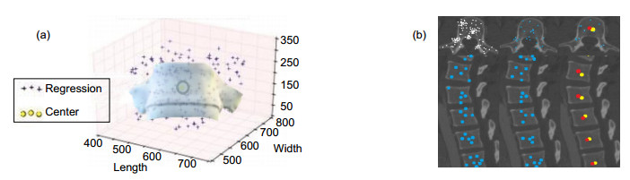
Figure 2.
Random forest center point positioning. (a) 3D distance map; (b) Regression points obtained by regression forest
-
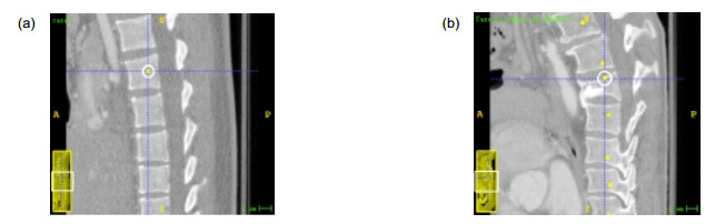
Figure 3.
Vertebral center and initial contours
-
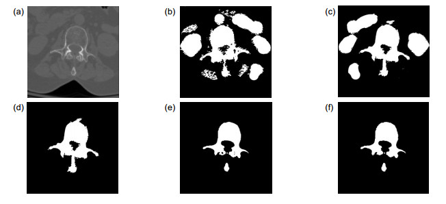
Figure 4.
Controlled variable experimental segmentation results
-
Figure 5.
Analysis of experimental segmentation results
-
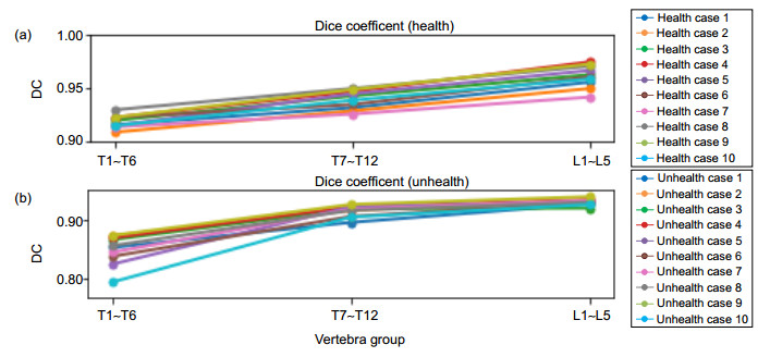
Figure 6.
DC and ASD statistical results
-
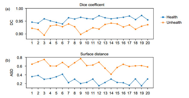
Figure 7.
DC coefficients and ASD coefficients of 20 cases
-
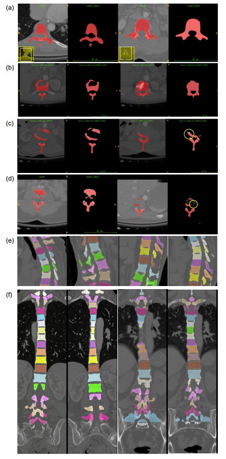
Figure 8.
Segmentation results of the proposed method
-
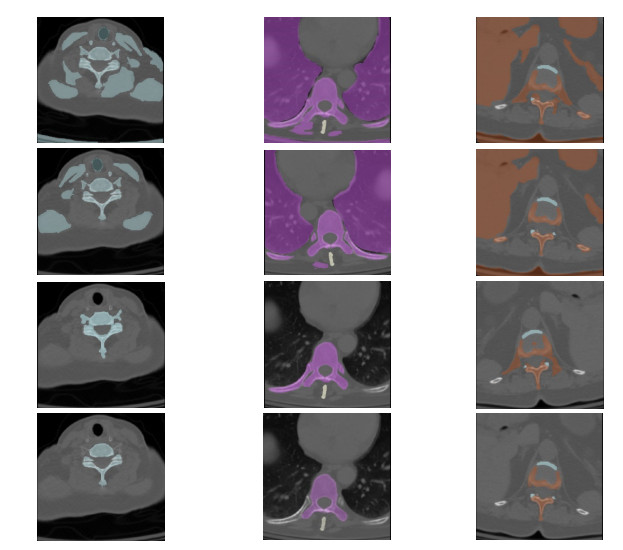
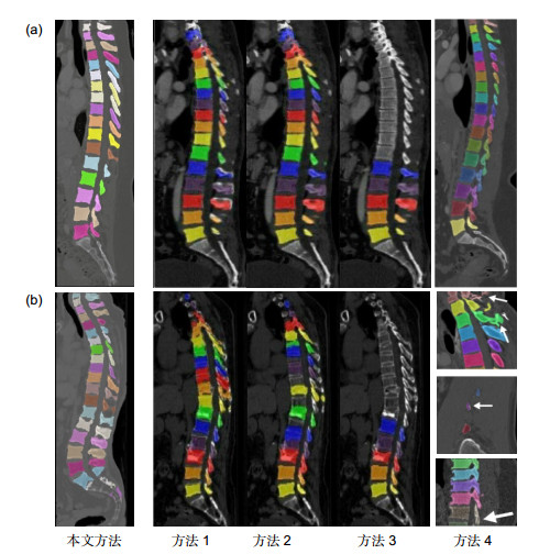
Figure 9.
Comparisons of the segmentation effect between the proposed method and other methods
-
Figure 10.
Visualization of 3D reconstruction of vertebrae


