
 E-mail Alert
 RSS
RSS
| Citation: |

|
The research progress and challenge of 3D bioprinting for skin repairing
-
Abstract
The skin is the largest organ of the human body, which plays an important role in barrier function, immune response, preventing water loss and excreting waste. Patients with large-scale severe skin injuries will die due to lack of adequate skin grafts. The development of 3D bioprinting technology provides a solution for the manufacture of transplantable skin. Firstly, the principles of skin wound repairing are described. Secondly, the bioinks, cells and main 3D bioprinting technologies used in skin wound repairing are compared. Then, the opto-electronic technologies involved are analyzed, and the challenges and future development of 3D bioprinting in the application of skin repair are summarized. Finally, the application requirements of opto-electronic technology in 3D bioprinting are proposed.-
Keywords:
- 3D bioprinting /
- skin /
- bioink /
- cell /
- opto-electronic technology
-

-
References
[1] Groeber F, Holeiter M, Hampel M, et al. Skin tissue engineering—In vivo and in vitro applications[J]. Adv Drug Deliv Rev, 2011, 63(4-5): 352-366. doi: 10.1016/j.addr.2011.01.005 [2] Vijayavenkataraman S, Lu W F, Fuh J Y H. 3D bioprinting of skin: a state-of-the-art review on modelling, materials, and processes[J]. Biofabrication, 2016, 8(3): 032001. doi: 10.1088/1758-5090/8/3/032001 [3] Peck M D. Epidemiology of burns throughout the World. Part Ⅱ: intentional burns in adults[J]. Burns, 2012, 38(5): 630-637. doi: 10.1016/j.burns.2011.12.028 [4] Beheshtizadeh N, Lotfibakhshaiesh N, Pazhouhnia Z, et al. A review of 3D bio-printing for bone and skin tissue engineering: a commercial approach[J]. J Mater Sci, 2019, 55(9): 3729-3749. doi: 10.1007/s10853-019-04259-0 [5] Derakhshanfar S, Mbeleck R, Xu K G, et al. 3D bioprinting for biomedical devices and tissue engineering: a review of recent trends and advances[J]. Bioact Mater, 2018, 3(2): 144-156. doi: 10.1016/j.bioactmat.2017.11.008 [6] Tan S H, Ngo Z H, Leavesley D, et al. Recent advances in the design of three-dimensional and bioprinted scaffolds for full-thickness wound healing[J]. Tissue Eng Part B: Rev, 2021, doi: 10.1089/ten.TEB.2020.0339. [7] Ozbolat I T. Bioprinting scale-up tissue and organ constructs for transplantation[J]. Trends Biotechnol, 2015, 33(7): 395-400. doi: 10.1016/j.tibtech.2015.04.005 [8] Chouhan D, Dey N, Bhardwaj N, et al. Emerging and innovative approaches for wound healing and skin regeneration: current status and advances[J]. Biomaterials, 2019, 216: 119267. doi: 10.1016/j.biomaterials.2019.119267 [9] Clark R A F, Ghosh K, Tonnesen M G. Tissue engineering for cutaneous wounds[J]. J Invest Dermatol, 2007, 127(5): 1018-1029. doi: 10.1038/sj.jid.5700715 [10] Rodrigues M, Kosaric N, Bonham C A, et al. Wound healing: a cellular perspective[J]. Physiol Rev, 2019, 99(1): 665-706. doi: 10.1152/physrev.00067.2017 [11] Yannas I V, Tzeranis D, So P T. Surface biology of collagen scaffold explains blocking of wound contraction and regeneration of skin and peripheral nerves[J]. Biomed Mater, 2015, 11(1): 014106. doi: 10.1088/1748-6041/11/1/014106 [12] Bhardwaj N, Chouhan D, Mandal B B. Tissue engineered skin and wound healing: current strategies and future directions[J]. Curr Pharm Des, 2017, 23(24): 3455-3482. doi: 10.2174/1381612823666170526094606 [13] Liu T, Qiu C, Ben C, et al. One-step approach for full-thickness skin defect reconstruction in rats using minced split-thickness skin grafts with Pelnac overlay[J]. Burns Trauma, 2019, 7: 19. doi: 10.1186/s41038-019-0157-0 [14] Haflah N H M, Ng M H, Yunus M H M, et al. Massive traumatic skin defect successfully treated with autologous, bilayered, tissue-engineered MyDerm skin substitute: a case report[J]. JBJS Case Connect, 2018, 8(2): e38. doi: 10.2106/JBJS.CC.17.00250 [15] Kirchmajer D M, Gorkin III R, in het Panhuis M. An overview of the suitability of hydrogel-forming polymers for extrusion-based 3D-printing[J]. J Mater Chem B, 2015, 3(20): 4105-4117. doi: 10.1039/C5TB00393H [16] Parak A, Pradeep P, du Toit L C, et al. Functionalizing bioinks for 3D bioprinting applications[J]. Drug Discov Today, 2019, 24(1): 198-205. doi: 10.1016/j.drudis.2018.09.012 [17] Agrawal P, Soni S, Mittal G, et al. Role of polymeric biomaterials as wound healing agents[J]. Int J Lower Extrem Wounds, 2014, 13(3): 180-190. doi: 10.1177/1534734614544523 [18] Gunatillake P A, Adhikari R. Biodegradable synthetic polymers for tissue engineering[J]. Eur Cells Mater, 2003, 5: 1-16. doi: 10.22203/eCM.v005a01 [19] Gugerell A, Pasteiner W, Nürnberger S, et al. Thrombin as important factor for cutaneous wound healing: comparison of fibrin biomatrices in vitro and in a rat excisional wound healing model[J]. Wound Repair Regen, 2014, 22(6): 740-748. doi: 10.1111/wrr.12234 [20] Currie L J, Martin R, Sharpe J R, et al. A comparison of keratinocyte cell sprays with and without fibrin glue[J]. Burns, 2003, 29(7): 677-685. doi: 10.1016/S0305-4179(03)00155-4 [21] Cubo N, Garcia M, del Cañizo J F, et al. 3D bioprinting of functional human skin: production and in vivo analysis[J]. Biofabrication, 2017, 9(1): 015006. doi: 10.1088/1758-5090/9/1/015006 [22] Donderwinkel I, van Hest J C M, Cameron N R. Bio-inks for 3D bioprinting: recent advances and future prospects[J]. Polym Chem, 2017, 8(31): 4451-4471. doi: 10.1039/C7PY00826K [23] Thiele J, Ma Y J, Bruekers S M C, et al. 25th anniversary article: designer hydrogels for cell cultures: a materials selection guide[J]. Adv Mater, 2014, 26(1): 125-148. doi: 10.1002/adma.201302958 [24] Kim Y B, Lee H, Kim G H. Strategy to achieve highly porous/biocompatible macroscale cell blocks, using a collagen/genipin-bioink and an optimal 3D printing process[J]. ACS Appl Mater Interfaces, 2016, 8(47): 32230-32240. doi: 10.1021/acsami.6b11669 [25] Lee J W, Choi Y J, Yong W J, et al. Development of a 3D cell printed construct considering angiogenesis for liver tissue engineering[J]. Biofabrication, 2016, 8(1): 015007. doi: 10.1088/1758-5090/8/1/015007 [26] Norouzi M, Boroujeni S M, Omidvarkordshouli N, et al. Advances in skin regeneration: application of electrospun scaffolds[J]. Adv Healthc Mater, 2015, 4(8): 1114-1133. doi: 10.1002/adhm.201500001 [27] Liu P C, Shen H Z, Zhi Y, et al. 3D bioprinting and in vitro study of bilayered membranous construct with human cells-laden alginate/gelatin composite hydrogels[J]. Colloids Surf B: Biointerfaces, 2019, 181: 1026-1034. doi: 10.1016/j.colsurfb.2019.06.069 [28] Del Amo C, Perez-Valle A, Perez-Zabala E, et al. Wound dressing selection is critical to enhance platelet-rich fibrin activities in wound care[J]. Int J Mol Sci, 2020, 21(2): 624. doi: 10.3390/ijms21020624 [29] Bociaga D, Bartniak M, Grabarczyk J, et al. Sodium alginate/gelatine hydrogels for direct bioprinting-the effect of composition selection and applied solvents on the bioink properties[J]. Materials, 2019, 12(17): 2669. doi: 10.3390/ma12172669 [30] Wang J, Shang P J, Shi W B, et al. Dissimilarity measure based on ordinal pattern for physiological signals[J]. Commun Nonlinear Sci Numer Simul, 2016, 37: 115-124. doi: 10.1016/j.cnsns.2016.01.011 [31] Pisani S, Dorati R, Scocozza F, et al. Preliminary investigation on a new natural based poly(gamma-glutamic acid)/Chitosan bioink[J]. J Biomed Mater Res Part B: Appl Biomater, 2020, 108(7): 2718-2732. doi: 10.1002/jbm.b.34602 [32] Li Y, Jiang X L, Li L, et al. 3D printing human induced pluripotent stem cells with novel hydroxypropyl chitin bioink: scalable expansion and uniform aggregation[J]. Biofabrication, 2018, 10(4): 044101. doi: 10.1088/1758-5090/aacfc3 [33] Dzobo K, Motaung K S C M, Adesida A. Recent trends in decellularized extracellular matrix bioinks for 3D printing: an updated review[J]. Int J Mol Sci, 2019, 20(18): 4628. doi: 10.3390/ijms20184628 [34] Kim B S, Kwon Y W, Kong J S, et al. 3D cell printing of in vitro stabilized skin model and in vivo pre-vascularized skin patch using tissue-specific extracellular matrix bioink: a step towards advanced skin tissue engineering[J]. Biomaterials, 2018, 168: 38-53. doi: 10.1016/j.biomaterials.2018.03.040 [35] Lee S J, Lee J H, Park J, et al. Fabrication of 3D printing scaffold with porcine skin decellularized bio-ink for soft tissue engineering[J]. Materials, 2020, 13(16): 3522. doi: 10.3390/ma13163522 [36] 吴兴, 刘肇兴, 林欢欢, 等. 甲基丙烯酸酐明胶材料学特性及在皮肤组织工程应用与进展[J]. 中国组织工程研究, 2018, 22(2): 323-328. doi: 10.3969/j.issn.2095-4344.0025 Wu X, Liu Z X, Lin H H, et al. Properties of gelatin methacryloyl and its application in the skin tissue engineering[J]. Chin J Tissue Eng Res, 2018, 22(2): 323-328. doi: 10.3969/j.issn.2095-4344.0025 [37] Zhao X, Lang Q, Yildirimer L, et al. Photocrosslinkable gelatin hydrogel for epidermal tissue engineering[J]. Adv Healthc Mater, 2016, 5(1): 108-118. doi: 10.1002/adhm.201500005 [38] Klotz B J, Gawlitta D, Rosenberg A J W P, et al. Gelatin-methacryloyl hydrogels: towards biofabrication-based tissue repair[J]. Trends Biotechnol, 2016, 34(5): 394-407. doi: 10.1016/j.tibtech.2016.01.002 [39] Bianco P, Robey P G. Stem cells in tissue engineering[J]. Nature, 2001, 414(6859): 118-121. doi: 10.1038/35102181 [40] Boyce S T, Medrano E E, Abdel-Malek Z, et al. Pigmentation and inhibition of wound contraction by cultured skin substitutes with adult melanocytes after transplantation to athymic mice[J]. J Invest Dermatol, 1993, 100(4): 360-365. doi: 10.1111/1523-1747.ep12471822 [41] Duncan C O, Shelton R M, Navsaria H, et al. In vitro transfer of keratinocytes: comparison of transfer from fibrin membrane and delivery by aerosol spray[J]. J Biomed Mater Res Part B: Appl Biomater, 2005, 73B(2): 221-228. doi: 10.1002/jbm.b.30198 [42] Sumorejo P, Listiawan M Y, Putri A I, et al. The role of stem cell metabolites derived from placenta for skin regeneration: an in vitro study[J]. Bali Med J, 2019, 8(1): 354-359. doi: 10.15562/bmj.v8i1.1387 [43] Mahmood R, Mehmood A, Choudhery M S, et al. Human neonatal stem cell-derived skin substitute improves healing of severe burn wounds in a rat model[J]. Cell Biol Int, 2019, 43(2): 147-157. doi: 10.1002/cbin.11072 [44] Metcalfe A D, Ferguson M W J. Skin stem and progenitor cells: using regeneration as a tissue-engineering strategy[J]. Cell Mol Life Sci, 2008, 65(1): 24-32. doi: 10.1007/s00018-007-7427-x [45] Lee W, Debasitis J C, Lee V K, et al. Multi-layered culture of human skin fibroblasts and keratinocytes through three-dimensional freeform fabrication[J]. Biomaterials, 2009, 30(8): 1587-1595. doi: 10.1016/j.biomaterials.2008.12.009 [46] Kim B S, Lee J S, Gao G, et al. Direct 3D cell-printing of human skin with functional transwell system[J]. Biofabrication, 2017, 9(2): 025034. doi: 10.1088/1758-5090/aa71c8 [47] Nolte S V, Xu W, Rennekampff H O, et al. Diversity of fibroblasts-a review on implications for skin tissue engineering[J]. Cells Tissues Organs, 2008, 187(3): 165-176. doi: 10.1159/000111805 [48] Won J Y, Lee M H, Kim M J, et al. A potential dermal substitute using decellularized dermis extracellular matrix derived bio-ink[J]. Artif Cells, Nanomed, Biotechnol, 2019, 47(1): 644-649. doi: 10.1080/21691401.2019.1575842 [49] Shi L, Xiong L M, Hu Y Q, et al. Three-dimensional printing alginate/gelatin scaffolds as dermal substitutes for skin tissue engineering[J]. Polym Eng Sci, 2018, 58(10): 1782-1790. doi: 10.1002/pen.24779 [50] Baltazar T, Merola J, Catarino C, et al. Three dimensional bioprinting of a vascularized and perfusable skin graft using human keratinocytes, fibroblasts, pericytes, and endothelial cells[J]. Tissue Eng Part A, 2020, 26(5-6): 227-238. doi: 10.1089/ten.tea.2019.0201 [51] Huyan Y G, Lian Q, Zhao T Z, et al. Pilot study of the biological properties and vascularization of 3D printed bilayer skin grafts[J]. Int J Bioprint, 2020, 6(1): 246. doi: 10.18063/ijb.v6i1.246 [52] Ng W L, Qi J T Z, Yeong W Y, et al. Proof-of-concept: 3D bioprinting of pigmented human skin constructs[J]. Biofabrication, 2018, 10(2): 025005. doi: 10.1088/1758-5090/aa9e1e [53] Min D, Lee W, Bae I H, et al. Bioprinting of biomimetic skin containing melanocytes[J]. Exp Dermatol, 2018, 27(5): 453-459. doi: 10.1111/exd.13376 [54] Dominici M, Le Blanc K, Mueller I, et al. Minimal criteria for defining multipotent mesenchymal stromal cells. The International Society for Cellular Therapy position statement[J]. Cytotherapy, 2006, 8(4): 315-317. doi: 10.1080/14653240600855905 [55] Kim H J, Park J S. Usage of human mesenchymal stem cells in cell-based therapy: advantages and disadvantages[J]. Dev Reprod, 2017, 21(1): 1-10. doi: 10.12717/DR.2017.21.1.001 [56] Kokubun K, Pankajakshan D, Kim M J, et al. Differentiation of porcine mesenchymal stem cells into epithelial cells as a potential therapeutic application to facilitate epithelial regeneration[J]. J Tissue Eng Regen Med, 2016, 10(2): E73-E83. doi: 10.1002/term.1758 [57] Strong A L, Neumeister M W, Levi B. Stem cells and tissue engineering: regeneration of the skin and its contents[J]. Clin Plastic Surg, 2017, 44(3): 635-650. doi: 10.1016/j.cps.2017.02.020 [58] Sasaki M, Abe R, Fujita Y, et al. Mesenchymal stem cells are recruited into wounded skin and contribute to wound repair by transdifferentiation into multiple skin cell type[J]. J Immunol, 2008, 180(4): 2581-2587. doi: 10.4049/jimmunol.180.4.2581 [59] Wu Y J, Chen L W, Scott P G, et al. Mesenchymal stem cells enhance wound healing through differentiation and angiogenesis[J]. Stem Cells, 2007, 25(10): 2648-2659. doi: 10.1634/stemcells.2007-0226 [60] Xiong J C, Ji B Y, Wang L J, et al. Human adipose-derived stem cells promote seawater-immersed wound healing by activating skin stem cells via the EGFR/MEK/ERK pathway[J]. Stem Cells Int, 2019, 2019: 7135974. doi: 10.1155/2019/7135974 [61] Visscher D O, Farré-Guasch E, Helder M N, et al. Advances in bioprinting technologies for craniofacial reconstruction[J]. Trends Biotechnol, 2016, 34(9): 700-710. doi: 10.1016/j.tibtech.2016.04.001 [62] Murphy S V, Atala A. 3D bioprinting of tissues and organs[J]. Nat Biotechnol, 2014, 32(8): 773-785. doi: 10.1038/nbt.2958 [63] Xu T, Jin J, Gregory C, et al. Inkjet printing of viable mammalian cells[J]. Biomaterials, 2005, 26(1): 93-99. doi: 10.1016/j.biomaterials.2004.04.011 [64] Park J A, Yoon S, Kwon J, et al. Freeform micropatterning of living cells into cell culture medium using direct inkjet printing[J]. Sci Rep, 2017, 7(1): 14610. doi: 10.1038/s41598-017-14726-w [65] Yoon S, Park J A, Lee H R, et al. Inkjet-spray hybrid printing for 3D freeform fabrication of multilayered hydrogel structures[J]. Adv Healthc Mater, 2018, 7(14): 1800050. doi: 10.1002/adhm.201800050 [66] Xu C X, Zhang M, Huang Y, et al. Study of droplet formation process during drop-on-demand inkjetting of living cell-laden bioink[J]. Langmuir, 2014, 30(30): 9130-9138. doi: 10.1021/la501430x [67] Negro A, Cherbuin T, Lutolf M P. 3D inkjet printing of complex, cell-laden hydrogel structures[J]. Sci Rep, 2018, 8(1): 17099. doi: 10.1038/s41598-018-35504-2 [68] Park J A, Lee H R, Park S Y, et al. Self-organization of fibroblast-laden 3D collagen microstructures from inkjet-printed cell patterns[J]. Adv Biosyst, 2020, 4(5): 1900280. doi: 10.1002/adbi.201900280 [69] Kim B S, Gao G, Kim J Y, et al. 3D cell printing of perfusable vascularized human skin equivalent composed of epidermis, dermis, and hypodermis for better structural recapitulation of native skin[J]. Adv Healthc Mater, 2019, 8(7): 1801019. doi: 10.1002/adhm.201801019 [70] Lim K S, Levato R, Costa P F, et al. Bio-resin for high resolution lithography-based biofabrication of complex cell-laden constructs[J]. Biofabrication, 2018, 10(3): 034101. doi: 10.1088/1758-5090/aac00c [71] Zhang A P, Qu X, Soman P, et al. Rapid fabrication of complex 3D extracellular microenvironments by dynamic optical projection stereolithography[J]. Adv Mater, 2012, 24(31): 4266-4270. doi: 10.1002/adma.201202024 [72] Ma X Y, Qu X, Zhu W, et al. Deterministically patterned biomimetic human iPSC-derived hepatic model via rapid 3D bioprinting[J]. Proc Natl Acad Sci USA, 2016, 113(8): 2206-2211. doi: 10.1073/pnas.1524510113 [73] Zhu W, Qu X, Zhu J, et al. Direct 3D bioprinting of prevascularized tissue constructs with complex microarchitecture[J]. Biomaterials, 2017, 124: 106-115. doi: 10.1016/j.biomaterials.2017.01.042 [74] Zhou F F, Hong Y, Liang R J, et al. Rapid printing of bio-inspired 3D tissue constructs for skin regeneration[J]. Biomaterials, 2020, 258: 120287. doi: 10.1016/j.biomaterials.2020.120287 [75] Hribar K C, Soman P, Warner J, et al. Light-assisted direct-write of 3D functional biomaterials[J]. Lab Chip, 2014, 14(2): 268-275. doi: 10.1039/C3LC50634G [76] Yan W C, Davoodi P, Vijayavenkataraman S, et al. 3D bioprinting of skin tissue: from pre-processing to final product evaluation[J]. Adv Drug Deliv Rev, 2018, 132: 270-295. doi: 10.1016/j.addr.2018.07.016 [77] Koch L, Kuhn S, Sorg H, et al. Laser printing of skin cells and human stem cells[J]. Tissue Eng Part C: Methods, 2010, 16(5): 847-854. doi: 10.1089/ten.tec.2009.0397 [78] Koch L, Deiwick A, Schlie S, et al. Skin tissue generation by laser cell printing[J]. Biotechnol Bioeng, 2012, 109(7): 1855-1863. doi: 10.1002/bit.24455 [79] Wang R, Wang Y H, Yao B, et al. Beyond 2D: 3D bioprinting for skin regeneration[J]. Int Wound J, 2019, 16(1): 134-138. doi: 10.1111/iwj.13003 [80] Mandrycky C, Wang Z J, Kim K, et al. 3D bioprinting for engineering complex tissues[J]. Biotechnol Adv, 2016, 34(4): 422-434. doi: 10.1016/j.biotechadv.2015.12.011 [81] Di Bella C, Duchi S, O'connell C D, et al. In situ handheld three-dimensional bioprinting for cartilage regeneration[J]. J Tissue Eng Regen Med, 2018, 12(3): 611-621. doi: 10.1002/term.2476 [82] Li X, Lian Q, Li D C, et al. Development of a robotic arm based hydrogel additive manufacturing system for in-situ printing[J]. Appl Sci, 2017, 7(1): 73. doi: 10.3390/app7010073 [83] Lee J, Kim K E, Bang S, et al. A desktop multi-material 3D bio-printing system with open-source hardware and software[J]. Int J Precis Eng Manuf, 2017, 18(4): 605-612. doi: 10.1007/s12541-017-0072-x [84] Masaeli E, Forster V, Picaud S, et al. Tissue engineering of retina through high resolution 3-dimentional inkjet bioprinting[J]. Biofabrication, 2020, 12(2): 025006. doi: 10.1088/1758-5090/ab4a20 [85] Gao Q, He Y, Fu J Z, et al. Fabrication of shape controllable alginate microparticles based on drop-on-demand jetting[J]. J Sol-Gel Sci Technol, 2016, 77(3): 610-619. doi: 10.1007/s10971-015-3890-2 [86] Kumar H, Kim K. Stereolithography 3D Bioprinting[J]. Methods Mol Biol, 2020, 2140: 93-108. [87] Warner J, Soman P, Zhu W, et al. Design and 3D printing of hydrogel scaffolds with fractal geometries[J]. ACS Biomater Sci Eng, 2016, 2(10): 1763-1770. doi: 10.1021/acsbiomaterials.6b00140 [88] Michael S, Sorg H, Peck C T, et al. Tissue engineered skin substitutes created by laser-assisted bioprinting form skin-like structures in the dorsal skin fold chamber in mice[J]. PLoS One, 2013, 8(3): e57741. doi: 10.1371/journal.pone.0057741 [89] Gauvin R, Chen Y C, Lee J W, et al. Microfabrication of complex porous tissue engineering scaffolds using 3D projection stereolithography[J]. Biomaterials, 2012, 33(15): 3824-3834. doi: 10.1016/j.biomaterials.2012.01.048 [90] Gou M L, Qu X, Zhu W, et al. Bio-inspired detoxification using 3D-printed hydrogel nanocomposites[J]. Nat Commun, 2014, 5: 3774. doi: 10.1038/ncomms4774 [91] Li J B, Han Y, Zhu J H. Influence equilibrium problem research based on common interests in mobile social network[C]//Proceedings of the 12th International Conference on Mobile Ad-Hoc and Sensor Networks, 2016: 271-278. [92] Schiele N R, Corr D T, Huang Y, et al. Laser-based direct-write techniques for cell printing[J]. Biofabrication, 2010, 2(3): 032001. doi: 10.1088/1758-5082/2/3/032001 [93] Liu X, Michael S, Bharti K, et al. A biofabricated vascularized skin model of atopic dermatitis for preclinical studies[J]. Biofabrication, 2020, 12(3): 035002. doi: 10.1088/1758-5090/ab76a1 [94] Lian Q, Li X, Li D C, et al. Path planning method based on discontinuous grid partition algorithm of point cloud for in situ printing[J]. Rapid Prototyping J, 2019, 25(3): 602-613. doi: 10.1108/RPJ-03-2018-0056 [95] Binder K W. In situ bioprintingi of the skin[D]. Winston-Salem: Wake Forest School, 2011: 1-443. [96] Hakimi N, Cheng R, Leng L, et al. Handheld skin printer: in situ formation of planar biomaterials and tissues[J]. Lab Chip, 2018, 18(10): 1440-1451. doi: 10.1039/C7LC01236E [97] Cheng R Y, Eylert G, Gariepy J M, et al. Handheld instrument for wound-conformal delivery of skin precursor sheets improves healing in full-thickness burns[J]. Biofabrication, 2020, 12(2): 025002. doi: 10.1088/1758-5090/ab6413 [98] Choi H S, Gibbs S L, Lee J H, et al. Targeted zwitterionic near-infrared fluorophores for improved optical imaging[J]. Nat Biotechnol, 2013, 31(2): 148-153. doi: 10.1038/nbt.2468 [99] Park G K, Kim S H, Kim K, et al. Dual-channel fluorescence imaging of hydrogel degradation and tissue regeneration in the brain[J]. Theranostics, 2019, 9(15): 4255-4264. doi: 10.7150/thno.35606 [100] Chen Y W, Zhang J M, Liu X, et al. Noninvasive in vivo 3D bioprinting[J]. Sci Adv, 2020, 6(23): eaba7406. doi: 10.1126/sciadv.aba7406 [101] Liu W J, Zhang Y S, Heinrich M A, et al. Rapid continuous multimaterial extrusion bioprinting[J]. Adv Mater, 2017, 29(3): 1604630. doi: 10.1002/adma.201604630 [102] Pi Q M, Maharjan S, Yan X, et al. Digitally tunable microfluidic bioprinting of multilayered cannular tissues[J]. Adv Mater, 2018, 30(43): 1706913. doi: 10.1002/adma.201706913 [103] Keriquel V, Guillemot F, Arnault I, et al. In vivo bioprinting for computer- and robotic-assisted medical intervention: preliminary study in mice[J]. Biofabrication, 2010, 2(1): 014101. doi: 10.1088/1758-5082/2/1/014101 [104] Albanna M, Binder K W, Murphy S V, et al. In situ bioprinting of autologous skin cells accelerates wound healing of extensive excisional full-thickness wounds[J]. Sci Rep, 2019, 9(1): 1856. doi: 10.1038/s41598-018-38366-w [105] 陈凯云, 谢晓芹, 叶佩青. 航空压气机叶片型面在线激光测量系统设计[J]. 制造技术与机床, 2004(8): 53-56. doi: 10.3969/j.issn.1005-2402.2004.08.014 Chen K Y, Xie X Q, Ye P Q. Design on on-line laser measurement system for vane of aero-engine compressor[J]. Manuf Technol Mach Tool, 2004(8): 53-56. doi: 10.3969/j.issn.1005-2402.2004.08.014 [106] Li L, Yu F, Shi J P, et al. In situ repair of bone and cartilage defects using 3D scanning and 3D printing[J]. Sci Rep, 2017, 7(1): 9416. doi: 10.1038/s41598-017-10060-3 [107] Atala A, Kasper F K, Mikos A G. Engineering complex tissues[J]. Sci Transl Med, 2012, 4(160): 160rv12. [108] Delrot P, Modestino M A, Gallaire F, et al. Inkjet printing of viscous monodisperse microdroplets by laser-induced flow focusing[J]. Phys Rev Appl, 2016, 6(2): 024003. doi: 10.1103/PhysRevApplied.6.024003 [109] Xue D, Wang Y C, Zhang J X, et al. Projection-based 3D printing of cell patterning scaffolds with multiscale channels[J]. ACS Appl Mater Interfaces, 2018, 10(23): 19428-19435. doi: 10.1021/acsami.8b03867 [110] Kattamis N T, Purnick P E, Weiss R, et al. Thick film laser induced forward transfer for deposition of thermally and mechanically sensitive materials[J]. Appl Phys Lett, 2007, 91(17): 171120. doi: 10.1063/1.2799877 [111] Guillotin B, Souquet A, Catros S, et al. Laser assisted bioprinting of engineered tissue with high cell density and microscale organization[J]. Biomaterials, 2010, 31(28): 7250-7256. doi: 10.1016/j.biomaterials.2010.05.055 [112] Zhu Z J, Guo S Z, Hirdler T, et al. 3D printed functional and biological materials on moving freeform surfaces[J]. Adv Mater, 2018, 30(23): 1707495. doi: 10.1002/adma.201707495 [113] Suh Y J, Lim T H, Choi H S, et al. 3D printing and NIR fluorescence imaging techniques for the fabrication of implants[J]. Materials, 2020, 13(21): 4819. doi: 10.3390/ma13214819 -
Overview

Overview: The skin is the first line of defense against external stimuli. Therefore, the skin is most vulnerable to injury, and serious skin injury may be life-threatening, so repairing damaged skin is of great significance. Because 3D bioprinting is able to accurately place a variety of different types of cells, even stem cells and appendages, and can repeatably create skin substitutes to replace the injured or damaged parts of the skin, making it similar to the skin appearance and function, 3D bioprinting makes up for the shortcomings of conventional skin wound repairing treatment, and is currently one of the most likely manufacturing methods to develop skin substitutes.
In order to improve the accuracy of printed skin, the degree of adaptation to the wound, and the effect of skin wound repairing, more and more opto-electronic technologies have been applied in 3D bioprinting. Piezoelectric and laser pulse technology can be used in the nozzle to obtain droplets with more uniform cell distribution and droplet diameters that are more suitable for inkjet printing. Digital mask projection technology uses digital micromirror to control the mask to print photosensitive materials to obtain high-resolution customized patterns. Laser-induced forward transfer technology adjusts the energy, spot size, and duration of the pulsed laser beam to cover the laser energy absorbing layer with bioink containing cells. Near-infrared fluorescence technology is used to monitor the printing process in real time, so as to adjust and plan the printing path in real time and obtain the skin with higher degree of compatibility with the defective skin.
In this article, firstly, the skin tissue structures and the principles of skin wound repairing are described. Secondly, the bioinks, cells and main 3D bioprinting technologies used in skin wound repairing are compared. Then, the opto-electronic technologies involved are analyzed. Finally, the application requirements of opto-electronic technology in 3D bioprinting are proposed.
-
Access History
Figures(4)
Tables(4)
Article Metrics

Export File
Citation
Format
Content
 DownLoad:

-
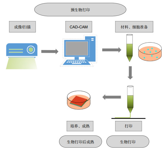
Figure 1.
Different steps and stages of 3D bioprinting
-
Figure 2.
Main process methods of 3D bioprinting for skin application.
-
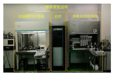
Figure 3.
Scanning and printing composite in-situ printing system
-
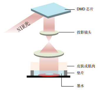
Figure 4.
Schematic diagram of DNP-based noninvasive 3D bioprinting[100]


