
 E-mail Alert
 RSS
RSS
| Citation: |

|
Optimized bow-tie metasurface and its application in trace detection of lead ion
-
Abstract
The strong localized plasmon resonance of metasurfaces makes the resonance frequency extremely sensitive to the dielectric environment, which can be applied to label-free environment detection. In this paper, a bow-tie terahertz metasurface with an optimized ratio of the quality factor to effective mode volume(Q/Veff) is designed. The unit cell of the proposed structure is composed of a mirror-symmetrical metallic bow tie in the middle and continuous metallic strips on both sides. The width of each metal strip and the length of the bow-tie gap are optimized for the parameter Q/Veff. When the metal strip width is 25 μm and the gap length is 2 μm, the effective mode volume is 3.6 μm3 and Q/Veff is 2.2 μm-3 at 0.7 THz. In the experiment, different concentration of the lead ion solution was dropped on the proposed metasurface. The transmission spectrum was measured by a terahertz time-domain spectroscopy system. The results showed that there is a linear relationship between resonance frequency shift and lead ion solution concentration from 0.1 ng/mL to 20 ng/mL. The detection limit is 0.1 ng/mL. The terahertz metasurface sensor has the advantages of the miniaturized size, easy sample preparation, fast measurement capability and real-time detection, which will be widely used in environmental protection and food safety.-
Keywords:
- terahertz /
- metasurface /
- lead ion /
- effective mode volume /
- high sensitivity
-

-
References
[1] Aragay G, Pons J, Merkoçi A. Recent trends in macro-, micro-, and nanomaterial-based tools and strategies for heavy-metal detection[J]. Chem Rev, 2011, 111(5): 3433-3458. doi: 10.1021/cr100383r [2] Dehouck P, Cordeiro F, Snell J, et al. State of the art in the determination of trace elements in seawater: a worldwide proficiency test[J]. Anal Bioanal Chem, 2016, 408(12): 3223-3232. doi: 10.1007/s00216-016-9390-6 [3] Fu F L, Wang Q. Removal of heavy metal ions from wastewaters: A review[J]. J Environ Manage, 2011, 92(3): 407-418. doi: 10.1016/j.jenvman.2010.11.011 [4] Wani A L, Ara A, Usmani J A. Lead toxicity: a review[J]. Interdiscip Toxicol, 2015, 8(2): 55-64. doi: 10.1515/intox-2015-0009 [5] Arora M, Kiran B, Rani S, et al. Heavy metal accumulation in vegetables irrigated with water from different sources[J]. Food Chem, 2008, 111(4): 811-815. doi: 10.1016/j.foodchem.2008.04.049 [6] Berlina A N, Zherdev A V, Dzantiev B B. Progress in rapid optical assays for heavy metal ions based on the use of nanoparticles and receptor molecules[J]. Microchim Acta, 2019, 186(3): 172. doi: 10.1007/s00604-018-3168-9 [7] Singh H, Bamrah A, Bhardwaj S K, et al. Nanomaterial-based fluorescent sensors for the detection of lead ions[J]. J Hazard Mater, 2021, 407: 124379. doi: 10.1016/j.jhazmat.2020.124379 [8] de Souza I D, de Andrade A S, Dalmolin R J S. Lead-interacting proteins and their implication in lead poisoning[J]. Crit Rev Toxicol, 2018, 48(5): 375-386. doi: 10.1080/10408444.2018.1429387 [9] Wallace D R, Djordjevic A B. Heavy metal and pesticide exposure: A mixture of potential toxicity and carcinogenicity[J]. Curr Opin Toxicol, 2020, 19: 72-79. doi: 10.1016/j.cotox.2020.01.001 [10] Acar O. Determination of cadmium, copper and lead in soils, sediments and sea water samples by ETAAS using a Sc + Pd + NHNO chemical modifier[J]. Talanta, 2005, 65(3): 672-677. doi: 10.1016/j.talanta.2004.07.035 [11] Koçak S, Tokuşoğlu Ö, Aycan Ş. Some heavy metal and trace essential element detection in canned vegetable foodstuffs by differential pulse polarography (DPP)[J]. Electronic J Environ Agric Food Chem, 2005, 4: 871-878. [12] Goossens J, Moens L, Dams R. Inductively coupled plasma mass spectrometric determination of heavy metals in soil and sludge candidate reference materials[J]. Anal Chim Acta, 1995, 304(3): 307-315. doi: 10.1016/0003-2670(94)00643-Z [13] Kot B, Baranowski R, Rybak A. Analysis of mine waters using X-ray fluorescence spectrometry[J]. Pol J Environ Stud, 2000, 9(5): 429-432. [14] Malitesta C, Guascito M R. Heavy metal determination by biosensors based on enzyme immobilised by electropolymerisation[J]. Biosens Bioelectron, 2005, 20(8): 1643-1647. doi: 10.1016/j.bios.2004.08.003 [15] De A, Kumari A, Jain P, et al. Plasmonic sensing of Hg(Ⅱ), Cr(Ⅲ), and Pb(Ⅱ) ions from aqueous solution by biogenic silver and gold nanoparticles[J]. Inorg Nano-Met Chem, 2021, 51(9): 1214-1225. [16] Sadrolhosseini A R, Shafie S, Rashid S A, et al. Surface plasmon resonance measurement of arsenic in low concentration using polypyrrole-graphene quantum dots layer[J]. Measurement, 2021, 173: 108546. doi: 10.1016/j.measurement.2020.108546 [17] Fen Y W, Yunus W M M, Yusof N A. Surface plasmon resonance optical sensor for detection of Pb2+ based on immobilized p-tert-butylcalix[4]arene-tetrakis in chitosan thin film as an active layer[J]. Sensor Actuat B: Chem, 2012, 171-172: 287-293. doi: 10.1016/j.snb.2012.03.070 [18] Chen L, Liao D G, Guo X G, et al. Terahertz time-domain spectroscopy and micro-cavity components for probing samples: a review[J]. Front Inform Technol Electron Eng, 2019, 20(5): 591-607. doi: 10.1631/FITEE.1800633 [19] Li B, Wang M H, Wang N, et al. Preliminary study on heavy metal detection in soil using terahertz time-domain spectroscopy[C]//2011 Louisville, Kentucky, August 7-10, 2011: 1100020(doi: 10.13031/2013.37181). [20] Li B, Wang M H, Cao W, et al. Research on heavy metal ions detection in soil with terahertz time-domain spectroscopy[J]. Proc SPIE, 2011, 8195: 81951V. doi: 10.1117/12.902302 [21] Lin S J, Xu X L, Hu F R, et al. Using antibody modified terahertz metamaterial biosensor to detect concentration of carcinoembryonic antigen[J]. IEEE J Sel Top Quantum Electron, 2020, 27(4): 6900207. [22] Yang Y P, Xu D Q, Zhang W L. High-sensitivity and label-free identification of a transgenic genome using a terahertz meta-biosensor[J]. Opt Express, 2018, 26(24): 31589-31598. doi: 10.1364/OE.26.031589 [23] Qin B Y, Li Z, Hu F R, et al. Highly sensitive detection of carbendazim by using terahertz time-domain spectroscopy combined with metamaterial[J]. IEEE Trans Terahertz Sci Technol, 2018, 8(2): 149-154. doi: 10.1109/TTHZ.2017.2787458 [24] Singh R, Cao W, Al-Naib I, et al. Ultrasensitive terahertz sensing with high-Q Fano resonances in metasurfaces[J]. Appl Phys Lett, 2014, 105(17): 171101. doi: 10.1063/1.4895595 [25] Zengin G, Wersäll M, Nilsson S, et al. Realizing strong light-matter interactions between single-nanoparticle plasmons and molecular excitons at ambient conditions[J]. Phys Rev Lett, 2015, 114(15): 157401. doi: 10.1103/PhysRevLett.114.157401 [26] Chen X X, Chen Y H, Qin J, et al. Mode modification of plasmonic gap resonances induced by strong coupling with molecular excitons[J]. Nano Lett, 2017, 17(5): 3246-3251. doi: 10.1021/acs.nanolett.7b00858 [27] Srinivasan K, Borselli M, Painter O, et al. Cavity Q, mode volume, and lasing threshold in small diameter AlGaAs microdisks with embedded quantum dots[J]. Opt Express, 2006, 14(3): 1094-1105. doi: 10.1364/OE.14.001094 [28] Gupta M, Singh R. Terahertz sensing with optimized Q/Veff metasurface cavities[J]. Adv Opt Mater, 2020, 8(16): 1902025. [29] Chen Y Q, Bi K X, Wang Q J, et al. Rapid focused ion beam milling based fabrication of plasmonic nanoparticles and assemblies via "Sketch and Peel" strategy[J]. ACS Nano, 2016, 10(12): 11228-11236. [30] Chen Y Q, Xiang Q, Li Z Q, et al. "Sketch and Peel" lithography for high-resolution multiscale patterning[J]. Nano Lett, 2016, 16(5): 3253-3259. [31] Hu J Y, Li Z, Zhai C Y, et al. Plasmonic photo-assisted electrochemical sensor for detection of trace lead ions based on Au anchored on two-dimensional g-C3N4/graphene nanosheets[J]. Rare Met, 2021, 40(7): 1727-1737. doi: 10.1007/s12598-020-01659-z -
Overview

Overview: Lead ion pollution in water is becoming a serious worldwide environmental problem. Effective and efficient detection of lead ion pollution requires sensitive and selective sensors with rapid on-site detection ability for performing the desired detection. Traditional spectroscopy, electrochemical, and inductively coupled plasma mass spectrometry (ICP-MS) methods for lead ion detection have some problems, such as complex experimental technology, long measuring period, expensive equipment and so on, which are only suitable for laboratory analysis. In this direction, one appropriate alternative approach is exploring plasmonic metasensor technology for low-level lead ion detection, which offers sophisticated opportunities in lead ion detection and ecological environment protection. Terahertz wave has unique characteristics of low photon energy, excellent security and the nonionizing effect. Metasurfaces are promising tools that have facilitated precise screening and recognition of diverse molecules and biomolecules through substantial field confinement at subwavelength geometries. The terahertz metasurface sensor stems from its capability to squeeze electromagnetic fields, simultaneously in frequency and space. However, the radiative and nonradiative losses limit the quality factor (Q) of the metasurface. The observation and study of the effectively low mode volume (Veff) were firstly reported in the middle of the twenty century, which was primarily established based on quantum electrodynamics. Metasurface designs with Q/Veff cavities become extremely important for enhancing the light-matter interaction.
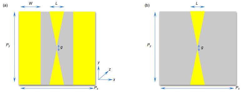
In this work, we demonstrate a modified bow-tie terahertz metasurface platform containing a micron-sized cavity with an optimized Q/Veff value. The structural unit is composed of a mirror symmetrical metallic bow tie in the middle and two continuous metallic strips on both sides. Continuous metallic strip enhances the overall capacitance of metasurface unit cell, which allows the capacitive split gap cavity to store larger electromagnetic energy. When the continuous metal strip width is 25 μm and the gap size is 2 μm, the Q/Veff of the proposed bow-tie terahertz metasurface reaches a maximum of 2.2 μm-3 at 0.7 THz. For 4 µm thick analyte layer, and the sensitivity is about 80 GHz/RIU (refractive index unit), which is higher than traditional bow tie metasurface. The proposed metasurface is manufactured using a surface micromachining process and characterized by a THz time-domain spectroscopy (THz-TDS) system. For the lead ion solution with different concentrations, the experimental results indicate that the resonance frequency of the terahertz metasurface sensor decreases with the increase of the concentration of lead ion solution on the surface. When the concentration of lead ion solution ranges from 0.1 ng/mL to 20 ng/mL, the resonance frequency shift shows good linearity to the concentration of lead ion solution and the limit of detection (LOD) reaches 0.1 ng/mL. It is expected to provide a new lead ion solution detection scheme for the field of environmental protection and food safety.
-
Access History

Export File
Citation
Format
Content
 DownLoad:
-
Figure 1.
(a) Top view of MBS design; (b) Top view of TBS design
-
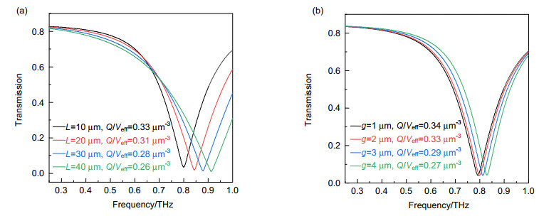
Figure 2.
(a) The effect of the bottom side L on transmission spectra and Q/Veff in TBS design; (b) The effect of the interval g on transmission spectra and Q/Veff in TBS design
-
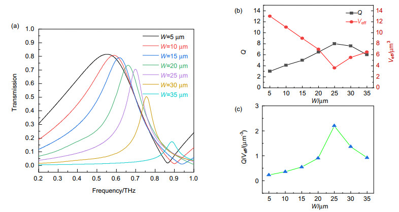
Figure 3.
(a) Transmission spectra with different values of the bandwidth W in MBS design; (b) The effect of the bandwidth W on the quality factor Q and effective mode volume Veff in MBS design; (c) The effect of the bandwidth W on Q/Veff in MBS design
-
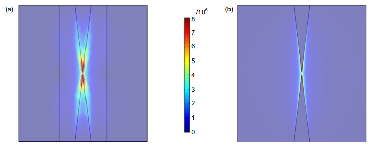
Figure 4.
Electric field distribution of metasurface at resonance frequencies.
-
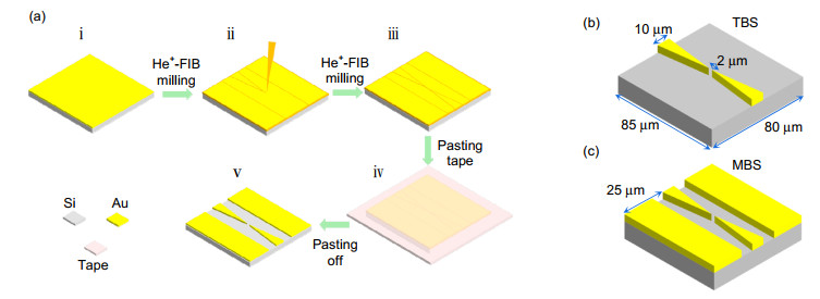
Figure 5.
(a) The fabrication flow chart of bow-tie metasurfaces by focused ion beam technology; (b) Stereogram of the TBS unit structure; (c) Stereogram of the MBS unit structure
-
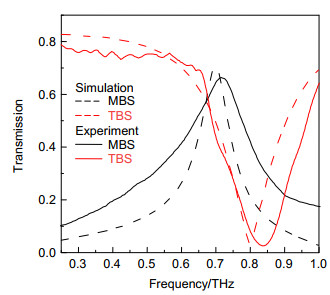
Figure 6.
Simulation and experimental transmission spectra of MBS and TBS designs
-
Figure 7.
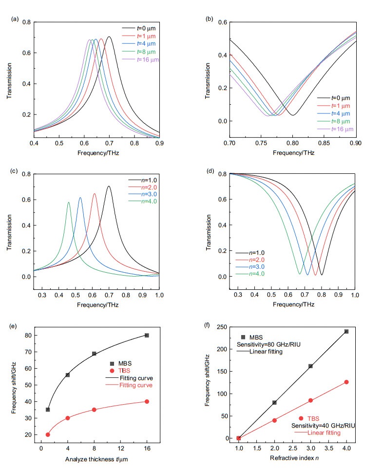
(a, b) Transmission spectra with respect to the thicknesses of analyte in (a) MBS design and (b) TBS design; (c, d) Transmission spectra with respect to refractive index in (c) MBS design and (d) TBS design; (e) Resonant peak shift with respect to the thicknesses of analyte in MBS and TBS design; (f) Resonant peak shift with respect to refractive index in MBS and TBS design
-
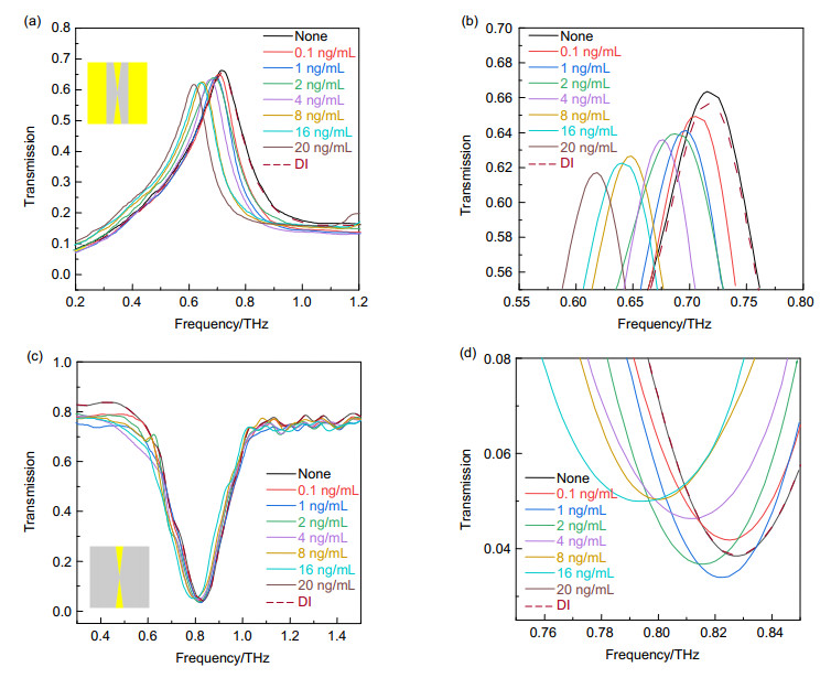
Figure 8.
(a) Transmission spectra of MBS for different concentrations of the lead ion solution; (b) Magnified view of Fig. 8(a); c) Transmission spectra of TBS for different concentrations of the lead ion solution; (d) Magnified view of Fig. 8(c)
-
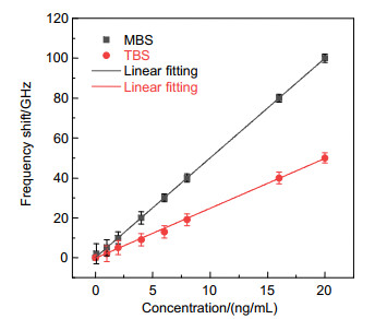
Figure 9.
The relationship between peak frequency shift of the metasurface sensor and concentrations of lead ion solution


