2023 Vol. 6, No. 1
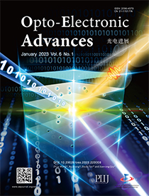
Cover story: Wang Z, Zhang B, Tan DZ, Qiu JR. Ostensibly perpetual optical data storage in glass with ultra-high stability and tailored photoluminescence. Opto-Electron Adv 6, 220008 (2023).
In the development history of human society, data storage plays an indispensable and pivotal role and has significantly boosted spacious domains from social science to industrial production. With the advent of the Internet of Things and artificial intelligence, the long lifetime and large capacity of information storage are in increasingly high demand. Recently, a team led by Prof. Jianrong Qiu from Zhejiang University and Prof. Dezhi Tan from Zhejiang Laboratory reported an ultra-long-term and high writing speed optical data storage technology enabled by single ultrafast laser pulse induced reduction of Eu3+ ions and tailoring of optical properties inside the Eu-doped aluminosilicate glasses. The lifetime of stored information is determined to be as long as twenty million years. The induced local modifications in the glass can stand against the temperature of up to 970 K and intense ultraviolet light irradiation with a power density of 100 kW/cm2. Furthermore, the active ions of Eu2+ exhibit strong and broadband emission with the full width at half maximum reaching 190 nm, and the photoluminescence is flexibly tunable in the whole visible region by regulating the alkaline earth metal ions in the glasses. The developed technology and materials will be of great significance in photonic applications such as long-term optical data storage.
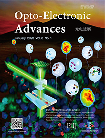
Back cover story: Pirone D, Sirico D, Miccio L, Bianco V, Mugnano M et al. 3D imaging lipidometry in single cell by in-flow holographic tomography. Opto-Electron Adv 6, 220048 (2023).
Scrutinizing a live cell in 3D can open novel perspective for full visualization and understanding the basic mechanism and function of intracellular structures. If this is made through a stain-free microscopy method and with the capability to achieve high-throughputs modality, this will be a game changing result in biology and clinics. The research group of Prof. Pietro Ferraro from National Research Council (CNR) of Italy in collaboration with the groups from University of Bologna has demonstrated for the first time the possibility to detect, visualize and quantify Lipids Droplets inside single live cell by holographic tomography combined with a microfluidic flow cytometry.
Investigation of LDs is an emergent field, especially since these organelles have been recognized as dynamic particles that play key roles in several cell regulation processes. In fact, it has been discovered that LDs number, size and interactions with other organelles reflect their functions.
Our approach demonstrates for the first time the capability to explore those parameters in a non-destructive, high-throughput mode and to furnish information of 3D spatial organization of Lipids Droplets (LDs) by tomography. So far, the techniques for study LDs have been transmission electron microscopy and fluorescent microscopy. However, even if such methods are highly specific and sensitive, they offer 2D analysis and on a small portion of the cell volume. Furthermore, both approaches are destructive and low throughput. Thus, the results reported in our work will be the first milestone along the path of the future development of flow cytometers for 3D analysis of label-free method that will offer much more information and data that could reveal the still unclear role of LDs.

-
{{article.year}}, {{article.volume}}({{article.issue}}): {{article.fpage | processPage:article.lpage:6}}. doi: {{article.doi}}{{article.articleStateNameEn}}, Published online {{article.preferredDate | date:'dd MMMM yyyy'}}, doi: {{article.doi}}{{article.articleStateNameEn}}, Accepted Date {{article.acceptedDate | date:'dd MMMM yyyy'}}CSTR: {{article.cstr}}

 E-mail Alert
 RSS
RSS


