
 E-mail Alert
 RSS
RSS
| Citation: |
Serien D, Sugioka K. Fabrication of three-dimensional proteinaceous micro- and nano-structures by femtosecond laser cross-linking. Opto-Electron Adv 1, 180008 (2018). doi: 10.29026/oea.2018.180008

|
Review Open Access
Fabrication of three-dimensional proteinaceous micro- and nano-structures by femtosecond laser cross-linking
-
Abstract
Proteins are a class of biomaterials having a vast array of functions, including the catalysis of metabolic reactions, DNA replication, stimuli response and transportation of molecules. Recent progress in laser-based fabrication technologies has enabled the formation of three-dimensional (3D) proteinaceous micro- and nano-structures by femtosecond laser cross-linking, which has expanded the possible applications of proteins. This article reviews the current knowledge and recent advancements in the femtosecond laser cross-linking of proteins. An overview of previous studies related to fabrication using a variety of proteins and detailed discussions of the associated mechanisms are provided. In addition, advances and applications utilizing specific protein functions are introduced. This review thus provides a valuable summary of the 3D micro- and nano-fabrication of proteins for biological and medical applications.-
Keywords:
- laser direct write /
- femtosecond laser /
- cross-linking /
- protein /
- 3D fabrication /
- microfluidics /
- actuation /
- scaffold
-

-
References
[1] Maskarinec S A, Tirrell D A. Protein engineering approaches to biomaterials design. Curr Opin Biotechnol 16, 422–426 (2005). doi: 10.1016/j.copbio.2005.06.009 [2] Latour Jr R A. Biomaterials: protein–surface interactions. In Bowlin G L, Wnek G. Encyclopedia of Biomaterials and Biomedical Engineering 270 (Marcel Dekker, 2013). [3] Ruel-Gariépy E, Leroux J C. In situ-forming hydrogels—review of temperature-sensitive systems. Eur J Pharm Biopharm 58, 409–426 (2004). doi: 10.1016/j.ejpb.2004.03.019 [4] Teixeira L S M, Feijen J, van Blitterswijk C A, Dijkstra P J, Karperien M. Enzyme-catalyzed crosslinkable hydrogels: emerging strategies for tissue engineering. Biomaterials 33, 1281–1290 (2012). doi: 10.1016/j.biomaterials.2011.10.067 [5] Shen W, Lammertink R G H, Sakata J K, Kornfield J A, Tirrell D A. Assembly of an artificial protein hydrogel through leucine zipper aggregation and disulfide bond formation. Macromolecules 38, 3909–3916 (2005). doi: 10.1021/ma048348s [6] Williams R J, Hall T E, Glattauer V, White J, Pasic P J et al. The in vivo performance of an enzyme-assisted self-assembled peptide/protein hydrogel. Biomaterials 32, 5304–5310 (2011). doi: 10.1016/j.biomaterials.2011.03.078 [7] Zubtsov D A, Ivanov S M, Rubina A Y, Dementieva E I, Chechetkin V R et al. Effect of mixing on reaction–diffusion kinetics for protein hydrogel-based microchips. J Biotechnol 122, 16–27 (2006). doi: 10.1016/j.jbiotec.2005.08.010 [8] Wu J H, Li P F, Dong C L, Jiang H T, Xue B et al. Rationally designed synthetic protein hydrogels with predictable mechanical properties. Nat Commun 9, 620 (2018). doi: 10.1038/s41467-018-02917-6 [9] Elzoghby A O, Samy W M, Elgindy N A. Protein-based nanocarriers as promising drug and gene delivery systems. J Control Release 161, 38–49 (2012). doi: 10.1016/j.jconrel.2012.04.036 [10] Kou S Z, Yang Z G, Sun F. Protein hydrogel microbeads for selective uranium mining from seawater. ACS Appl Mater Interfaces 9, 2035–2039 (2017). doi: 10.1021/acsami.6b15968 [11] Onoe H, Okitsu T, Itou A, Kato-Negishi M, Gojo R et al. Metre-long cell-laden microfibres exhibit tissue morphologies and functions. Nat Mater 12, 584–590 (2013). doi: 10.1038/nmat3606 [12] Hsiao A Y, Okitsu T, Onoe H, Kiyosawa M, Teramae H et al. Smooth muscle-like tissue constructs with circumferentially oriented cells formed by the cell fiber technology. PLoS One 10, e0119010 (2015). doi: 10.1371/journal.pone.0119010 [13] Kato-Negishi M, Onoe H, Ito A, Takeuchi S. Rod-shaped neural units for aligned 3D neural network connection. Adv Healthc Mater 6, 1700143 (2017). doi: 10.1002/adhm.v6.15 [14] Bahukudumbi P, Carson K H, Rice-Ficht A C, Andrews M J. On the diameter and size distributions of bovine serum albumin (BSA)-based microspheres. J Microencapsul 21, 787–803 (2004). doi: 10.1080/02652040400015395 [15] Suslick K S, Grinstaff M W, Kolbeck K J, Wong M. Characterization of sonochemically prepared proteinaceous microspheres. Ultrason Sonochem 1, S65-S68 (1994). doi: 10.1016/1350-4177(94)90030-2 [16] Silva R, Ferreira H, Vasconcelos A, Gomes A C, Cavaco-Paulo A. Sonochemical proteinaceous microspheres for wound healing. In Zahavy E, Ordentlich A, Yitzhaki S, Shafferman A. Nano-Biotechnology for Biomedical and Diagnostic Research 733 (Springer, 2012). [17] Forsberg F, Goldberg B B, Liu J B, Merton D A, Rawool N M. On the feasibility of real-time, in vivo harmonic imaging with proteinaceous microspheres. Ultrasound 15, 853–860 (1996). [18] Boland T, Xu T, Damon B, Cui X F. Application of inkjet printing to tissue engineering. Biotechnol J 1, 910–917 (2006). doi: 10.1002/(ISSN)1860-7314 [19] Roth E A, Xu T, Das M, Gregory C, Hickman J J et al. Inkjet printing for high-throughput cell patterning. Biomaterials 25, 3707–3715 (2004). doi: 10.1016/j.biomaterials.2003.10.052 [20] Geckil H, Xu F, Zhang X H, Moon S J, Utkan D. Engineering hydrogels as extracellular matrix mimics. Nanomedicine 5, 469–484 (2010). doi: 10.2217/nnm.10.12 [21] Kang H W, Lee S J, Ko I K, Kengla C, Yoo J J et al. A 3D bioprinting system to produce human-scale tissue constructs with structural integrity. Nat Biotechnol 34, 312–319 (2016). doi: 10.1038/nbt.3413 [22] Delaporte P, Alloncle A P. Laser-induced forward transfer: a high resolution additive manufacturing technology. Opt Laser Technol 78, 33–41 (2016). doi: 10.1016/j.optlastec.2015.09.022 [23] Zergioti I, Karaiskou A, Papazoglou D G, Fotakis C, Kapsetaki M et al. Femtosecond laser microprinting of biomaterials. Appl Phys Lett 86, 163902 (2005). doi: 10.1063/1.1906325 [24] Hopp B, Smausz T, Kresz N, Barna N, Bor Z et al. Survival and proliferative ability of various living cell types after laser-induced forward transfer. Tissue Eng 11, 1817–1823 (2005). doi: 10.1089/ten.2005.11.1817 [25] Kattamis N T, Purnick P E, Weiss R, Arnold C B. Thick film laser induced forward transfer for deposition of thermally and mechanically sensitive materials. Appl Phys Lett 91, 171120 (2007). doi: 10.1063/1.2799877 [26] Spikes J D, Shen H R, Kopečková P, Kopeček J. Photodynamic crosslinking of proteins. Ⅲ. Kinetics of the FMN-and rose Bengal-sensitized photooxidation and intermolecular crosslinking of model tyrosine-containing N-(2-hydroxypropyl) methacrylamide copolymers. Photochem Photobiol 70, 130–137 (1999). doi: 10.1111/php.1999.70.issue-2 [27] Moss T, Dimitrov S I, Houde D. UV-laser crosslinking of proteins to DNA. Methods 11, 225–234 (1997). doi: 10.1006/meth.1996.0409 [28] Gebicki S, Gebicki J M. Crosslinking of DNA and proteins induced by protein hydroperoxides. Biochem J 338, 629–636 (1999). doi: 10.1042/bj3380629 [29] Sugioka K, Cheng Y. Ultrafast lasers—reliable tools for advanced materials processing. Light Sci Appl 3, e149 (2014). doi: 10.1038/lsa.2014.30 [30] Sugioka K, Cheng Y. Femtosecond laser three-dimensional micro- and nanofabrication. Appl Phys Rev 1, 041303 (2014). doi: 10.1063/1.4904320 [31] Fischer J, Wegener M. Three-dimensional optical laser lithography beyond the diffraction limit. Laser Photon Rev 7, 22–44 (2013). doi: 10.1002/lpor.2013.7.issue-1 [32] Pitts J D, Campagnola P J, Epling G A, Goodman S L. Submicron multiphoton free-form fabrication of proteins and polymers: studies of reaction efficiencies and applications in sustained release. Macromolecules 33, 1514–1523 (2000). doi: 10.1021/ma9910437 [33] Serien D, Takeuchi S. Fabrication of submicron proteinaceous structures by direct laser writing. Appl Phys Lett 107, 013702 (2015). doi: 10.1063/1.4926659 [34] Spivey E C, Ritschdorff E T, Connell J L, McLennon C A, Schmidt C E et al. Multiphoton lithography of unconstrained three-dimensional protein microstructures. Adv Func Mater 23, 333–339 (2013). doi: 10.1002/adfm.201201465 [35] Lin C L, Pan M J, Chen H W, Lin C K, Lin C F et al. Laser cross-linking protein captures for living cells on a biochip. Proc SPIE 9310, 93100D (2015). [36] Iosin M, Scheul T, Nizak C, Stephan O, Astilean S et al. Laser microstructuration of three-dimensional enzyme reactors in microfluidic channels. Microfluid Nanofluid 10, 685–690 (2011). doi: 10.1007/s10404-010-0698-9 [37] Serien D, Kawano H, Miyawaki A, Midorikawa K, Sugioka K. Femtosecond laser direct write integration of multi-protein patterns and 3D microstructures into 3D glass microfluidic devices. Appl Sci 8, 147 (2018). doi: 10.3390/app8020147 [38] Basu S, Campagnola P J. Enzymatic activity of alkaline phosphatase inside protein and polymer structures fabricated via multiphoton excitation. Biomacromolecules 5, 572–579 (2004). doi: 10.1021/bm0344194 [39] Kaehr B, Shear J B. Multiphoton fabrication of chemically responsive protein hydrogels for microactuation. Proc Natl Acad Sci USA 105, 8850–8854 (2008). doi: 10.1073/pnas.0709571105 [40] Lee M R, Phang I Y, Cui Y, Lee Y H, Ling X Y. Shape-shifting 3D protein microstructures with programmable directionality via quantitative nanoscale stiffness modulation. Small 11, 740–748 (2015). doi: 10.1002/smll.201401343 [41] Sun Y L, Dong W F, Niu L G, Jiang T, Liu D X et al. Protein-based soft micro-optics fabricated by femtosecond laser direct writing. Light Sci Appl 3, e129 (2014). doi: 10.1038/lsa.2014.10 [42] Sun S M, Sun Y L, Zheng B Y, Wang P, Hou Z S et al. Protein-based Y-junction optical micro-splitters with environment-stimulus-actuated adjustments. Sens Actuators B Chem 232, 571–576 (2016). doi: 10.1016/j.snb.2016.03.164 [43] Serien D, Takeuchi S. Multi-Component microscaffold with 3D spatially defined proteinaceous environment. ACS Biomater Sci Eng 3, 487–494 (2017). doi: 10.1021/acsbiomaterials.6b00695 [44] Engelhardt S, Hoch E, Borchers K, Meyer W, Krüger H et al. Fabrication of 2D protein microstructures and 3D polymer–protein hybrid microstructures by two-photon polymerization. Biofabrication 3, 025003 (2011). doi: 10.1088/1758-5082/3/2/025003 [45] Khripin C Y, Brinker C J, Kaehr B. Mechanically tunable multiphoton fabricated protein hydrogels investigated using atomic force microscopy. Soft Matter 6, 2842–2848 (2010). doi: 10.1039/c001193b [46] Hill R T, Lyon J L, Allen R, Stevenson K J, Shear J B. Microfabrication of three-dimensional bioelectronic architectures. J Am Chem Soc 127, 10707–10711 (2005). doi: 10.1021/ja052211f [47] Connell J L, Ritschdorff E T, Shear J B. Three-dimensional printing of photoresponsive biomaterials for control of bacterial microenvironments. Anal Chem 88, 12264–12271 (2016). doi: 10.1021/acs.analchem.6b03440 [48] Turunen S, Käpylä E, Terzaki K, Viitanen J, Fotakis C et al. Pico- and femtosecond laser-induced crosslinking of protein microstructures: evaluation of processability and bioactivity. Biofabrication 3, 045002 (2011). doi: 10.1088/1758-5082/3/4/045002 [49] Kaehr B, Ertas N, Nielson R, Allen R, Hill R T et al. Direct-write fabrication of functional protein matrixes using a low-cost Q-switched laser. Anal Chem 78, 3198–3202 (2006). doi: 10.1021/ac052267s [50] Iosin M, Stephan O, Astilean S, Duperray A, Baldeck P. Microstructuration of protein matrices by laser-induced photochemistry. J Optoelectron Adv M 9, 716–720 (2007). [51] Basu S, Wolgemuth C W, Campagnola P J. Measurement of normal and anomalous diffusion of dyes within protein structures fabricated via multiphoton excited cross-linking. Biomacromolecules 5, 2347–2357 (2004). doi: 10.1021/bm049707u [52] Basu S, Cunningham L P, Pins G D, Bush K A, Taboada R et al. Multiphoton excited fabrication of collagen matrixes cross-linked by a modified benzophenone dimer: bioactivity and enzymatic degradation. Biomacromolecules 6, 1465–1474 (2005). doi: 10.1021/bm049258y [53] Sun Y L, Hou Z S, Sun S M, Zheng B Y, Ku J F et al. Protein-based three-dimensional whispering-gallery-mode micro-lasers with stimulus-responsiveness. Sci Rep 5, 12852 (2015). doi: 10.1038/srep12852 [54] Lawson J L, Jenness N, Wilson S, Clark R L. Method of creating microscale prototypes using SLM based holographic lithography. Proc SPIE 8612, 86120L (2013). [55] Sun Y L, Dong W F, Yang R Z, Meng X, Zhang L et al. Dynamically tunable protein microlenses. Angew Chem Int Ed 51, 1558–1562 (2012). doi: 10.1002/anie.v51.7 [56] Kaehr B, Allen R, Javier D J, Currie J, Shear J B. Guiding neuronal development with in situ microfabrication. Proc Natl Acad Sci USA 101, 16104–16108 (2004). doi: 10.1073/pnas.0407204101 [57] Harper J C, Brozik S M, Brinker C J, Kaehr B. Biocompatible microfabrication of 3D isolation chambers for targeted confinement of individual cells and their progeny. Anal Chem 84, 8985–8989 (2012). doi: 10.1021/ac301816c [58] Nielson R, Kaehr B, Shear J B. Microreplication and design of biological architectures using dynamic-mask multiphoton lithography. Small 5, 120–125 (2009). doi: 10.1002/smll.v5:1 [59] Da Sie Y, Li Y C, Chang N S, Campagnola P J, Chen S J. Fabrication of three-dimensional multi-protein microstructures for cell migration and adhesion enhancement. Biomed Opt Express 6, 480–490 (2015). doi: 10.1364/BOE.6.000480 [60] Ritschdorff E T, Nielson R, Shear J B. Multi-focal multiphoton lithography. Lab Chip 12, 867–871 (2012). doi: 10.1039/c2lc21271d [61] Allen R, Nielson R, Wise D D, Shear J B. Catalytic three-dimensional protein architectures. Anal Chem 77, 5089–5095 (2005). doi: 10.1021/ac0507892 [62] Lay C L, Lee M R, Lee H K, Phang I Y, Ling X Y. Transformative two-dimensional array configurations by geometrical shape-shifting protein microstructures. ACS Nano 9, 9708–9717 (2015). doi: 10.1021/acsnano.5b04300 [63] Lay C L, Lee Y H, Lee M R, Phang I Y, Ling X Y. Formulating an ideal protein photoresist for fabricating dynamic microstructures with high aspect ratios and uniform responsiveness. ACS Appli Mater Interfaces 8, 8145–8153 (2016). doi: 10.1021/acsami.6b02306 [64] Hernandez D S, Ritschdorff E T, Seidlits S K, Schmidt C E, Shear J B. Functionalizing micro-3D-printed protein hydrogels for cell adhesion and patterning. J Mater Chem B 4, 1818–1826 (2016). doi: 10.1039/C5TB02070K [65] Jenness N J, Hill R T, Hucknall A, Chilkoti A, Clark R L. A versatile diffractive maskless lithography for single-shot and serial microfabrication. Opt Express 18, 11754–11762 (2010). doi: 10.1364/OE.18.011754 [66] Serien D, Takeuchi S. Two-Photon direct laser writing for proteinaceous microstructures with additional sensitizer. J Laser Micro/Nanoeng 12, 80–85 (2017). doi: 10.2961/jlmn.2017.02.0006 [67] Bell A, Kofron M, Nistor V. Multiphoton crosslinking for biocompatible 3D printing of type I collagen. Biofabrication 7, 035007 (2015). doi: 10.1088/1758-5090/7/3/035007 [68] Lyon J L, Hill R T, Shear J B, Stevenson K J. Direct electrochemical and spectroscopic assessment of heme integrity in multiphoton photo-cross-linked cytochrome c structures. Anal Chem 79, 2303–2311 (2007). doi: 10.1021/ac0619377 [69] Basu S, Campagnola P J. Properties of crosslinked protein matrices for tissue engineering applications synthesized by multiphoton excitation. J Biomed Mater Res A 71A, 359–368 (2004). doi: 10.1002/(ISSN)1097-4636 [70] Sun Y L, Li Q, Sun S M, Huang J C, Zheng B Y et al. Aqueous multiphoton lithography with multifunctional silk-centred bio-resists. Nat Commun 6, 8612 (2015). doi: 10.1038/ncomms9612 [71] Vagenende V, Yap M G S, Trout B L. Mechanisms of protein stabilization and prevention of protein aggregation by glycerol. Biochemistry 48, 11084–11096 (2009). doi: 10.1021/bi900649t [72] Hand D B. The refractivity of protein solutions. J Biol Chem 108, 703–707 (1935). [73] Burley S K, Berman H M, Christie C, Duarte J M, Feng Z et al. RCSB Protein Data Bank: Sustaining a living digital data resource that enables breakthroughs in scientific research and biomedical education. Protein Sci 27, 316-330 (2018). doi: 10.1002/pro.3331 [74] Livingstone C D, Barton G J. Protein sequence alignments: a strategy for the hierarchical analysis of residue conservation. Bioinformatics 9, 745–756 (1993). doi: 10.1093/bioinformatics/9.6.745 [75] Eitner K, Koch U, Gawęda T, Marciniak J. Statistical distribution of amino acid sequences: a proof of Darwinian evolution. Bioinformatics 26, 2933–2935 (2010). doi: 10.1093/bioinformatics/btq571 [76] Peters T. Serum albumin. Adv Clin Chem 13, 37–111 (1970). doi: 10.1016/S0065-2423(08)60385-6 [77] Yu T, Ober C K, Kuebler S M, Zhou W, Marder S R et al. Chemically amplified positive resists for two-photon three-dimensional microfabrication. Adv Mater 15, 517–521 (2003). doi: 10.1002/adma.200390120 [78] Coenjarts C A, Ober C K. Two-photon three-dimensional microfabrication of poly(dimethylsiloxane) elastomers. Chem Mater 16, 5556–5558 (2004). doi: 10.1021/cm048717z [79] Oster G. Dye-sensitized photopolymerization. Nature 173, 300–301 (1954). doi: 10.1038/173300a0 [80] Dubbelman T M A R, De Goeij A F P M, Van Steveninck J. Protoporphyrin-induced photodynamic effects on transport processes across the membrane of human erythrocytes. Biochim Biophys Acta Biomembr 595, 133–139 (1980). doi: 10.1016/0005-2736(80)90255-2 [81] Shen H R, Spikes J D, Kopečková P, Kopeček J. Photodynamic crosslinking of proteins Ⅱ. Photocrosslinking of a model protein-ribonuclease A. J Photochem Photobiol B 35, 213–219 (1996). doi: 10.1016/S1011-1344(96)07300-9 [82] Requejo R, Hurd T R, Costa N J, Murphy M P. Cysteine residues exposed on protein surfaces are the dominant intramitochondrial thiol and may protect against oxidative damage. FEBS J 277, 1465–1480 (2010). doi: 10.1111/j.1742-4658.2010.07576.x [83] Sackmann E K, Fulton A L, Beebe D J. The present and future role of microfluidics in biomedical research. Nature 507, 181–189 (2014). doi: 10.1038/nature13118 [84] Huh D, Torisawa Y S, Hamilton G A, Kim H J, Ingber D E. Microengineered physiological biomimicry: organs-on-Chips. Lab Chip 12, 2156–2164 (2012). doi: 10.1039/c2lc40089h [85] Fan X D, White I M. Optofluidic microsystems for chemical and biological analysis. Nat Photonics 5, 591–597 (2011). doi: 10.1038/nphoton.2011.206 [86] Wu D, Wu S Z, Xu J, Niu L G, Midorikawa K et al. Hybrid femtosecond laser microfabrication to achieve true 3D glass/polymer composite biochips with multiscale features and high performance: the concept of ship-in-a-bottle biochip. Laser Photon Rev 8, 458–467 (2014). doi: 10.1002/lpor.201400005 -
Access History
Figures(8)
Tables(2)
Article Metrics

Export File
Citation
Serien D, Sugioka K. Fabrication of three-dimensional proteinaceous micro- and nano-structures by femtosecond laser cross-linking. Opto-Electron Adv 1, 180008 (2018). doi: 10.29026/oea.2018.180008
Format
Content
 DownLoad:
-
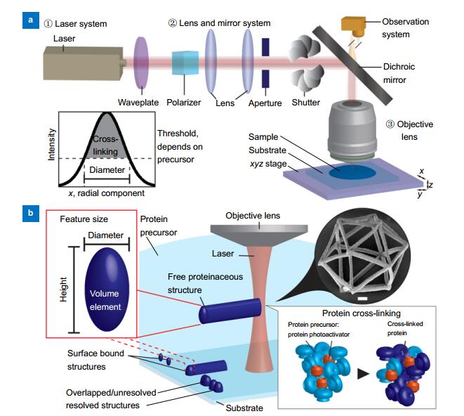
Figure 1.
Laser writing system for microfabrication of proteinaceous structures.
-
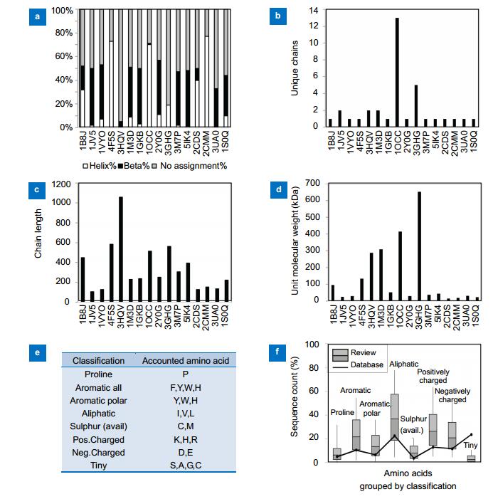
Figure 2.
PDB data overview.
-
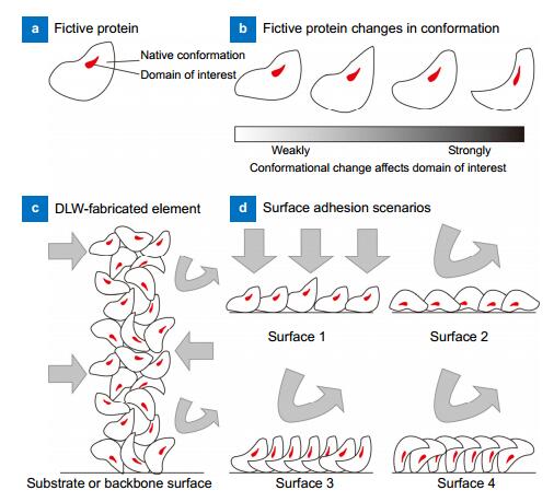
Figure 3.
Proposed processes for protein cross-linking and adherence.
-
Figure 4.
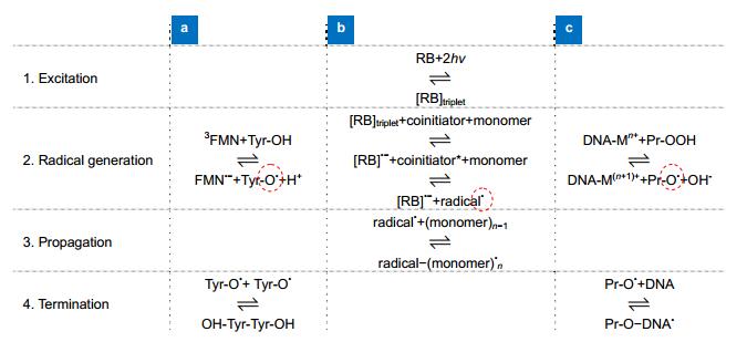
Proposed free radical mechanisms.
-
Figure 5.
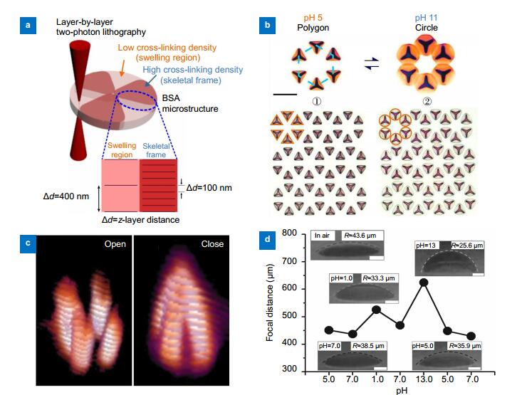
pH actuation.
-
Figure 6.
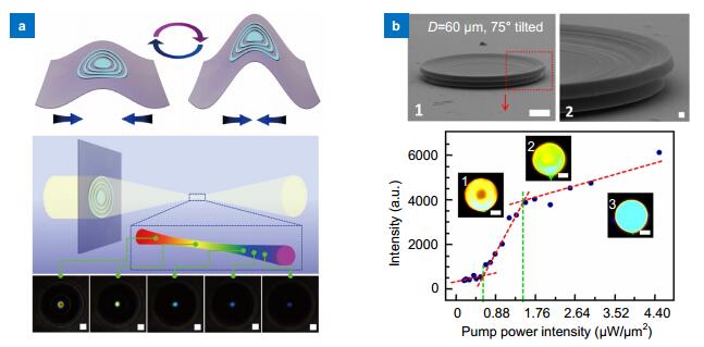
Soft microoptics.
-
Figure 7.
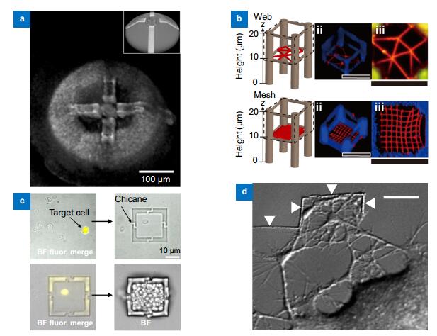
Cell culture scaffold and in situ guidance.
-
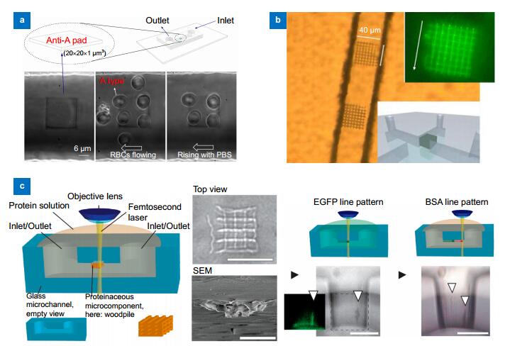
Figure 8.
Microfluidic integration.


