
 E-mail Alert
 RSS
RSS
| Citation: |
Yang LY, Li YP, Fang F, Li LY, Yan ZJ et al. Highly sensitive and miniature microfiber-based ultrasound sensor for photoacoustic tomography. Opto-Electron Adv 5, 200076 (2022). doi: 10.29026/oea.2022.200076

|
Highly sensitive and miniature microfiber-based ultrasound sensor for photoacoustic tomography
-
Abstract
A microfiber with large evanescent field encapsulated in PDMS is proposed and demonstrated for ultrasound sensing. The compact size and large evanescent field of microfiber provide an excellent platform for the interaction between optical signal and ultrasound wave, exhibiting a high sensitivity of 3.5 mV/kPa, which is approximately 10 times higher than the single-mode fiber sensor. Meanwhile, a phase feedback stabilization module is introduced into the coherent demodulation system for long-term stable measurement. In addition, a photoacoustic tomography experiment with the microfiber ultrasound sensor is implemented to verify the excellent performance on imaging, with the depth of 12 mm, the highest lateral resolution of 65 μm and axial resolution of 250 μm, respectively. The highly sensitive microfiber ultrasound sensor provides a competitive alternative for various applications, such as industrial non-destructive testing, biomedical ultrasound and photoacoustic imaging.-
Keywords:
- ultrasound sensor /
- microfiber /
- photoacoustic tomography
-

-
References
[1] Li C, Wang LV. Photoacoustic tomography and sensing in biomedicine. Phys Med Biol 54, R59–97 (2009). doi: 10.1088/0031-9155/54/19/R01 [2] Beard P. Biomedical photoacoustic imaging. Interface Focus 1, 602–631 (2011). doi: 10.1098/rsfs.2011.0028 [3] Powers J, Kremkau F. Medical ultrasound systems. Interface focus 1, 477–489 (2011). doi: 10.1098/rsfs.2011.0027 [4] Le Jeune L, Robert S, Villaverde EL, Prada C. Plane Wave Imaging for ultrasonic non-destructive testing: Generalization to multimodal imaging. Ultrasonics 64, 128–138 (2016). doi: 10.1016/j.ultras.2015.08.008 [5] Drinkwater BW, Wilcox PD. Ultrasonic arrays for non-destructive evaluation: A review. NDT E Int 39, 525–541 (2006). doi: 10.1016/j.ndteint.2006.03.006 [6] Hekmati A, Hekmati R. Optimum acoustic sensor placement for partial discharge allocation in transformers. IET Sci Meas Technol 11, 581–589 (2017). doi: 10.1049/iet-smt.2016.0417 [7] Sarkar B, Mishra DK, Koley C, Roy NK, Biswas P. Intensity-Modulated Fiber Bragg Grating Sensor for Detection of Partial Discharges Inside High-Voltage Apparatus. IEEE Sens J 16, 7950–7957 (2016). doi: 10.1109/JSEN.2016.2608743 [8] Janapati V, Kopsaftopoulos F, Li F, Lee SJ, Chang FK. Damage detection sensitivity characterization of acousto-ultrasound-based structural health monitoring techniques. Structural Health Monitoring-an International Journal 15, 143–161 (2016). doi: 10.1177/1475921715627490 [9] Qiu YQ, Gigliotti JV, Wallace M, Griggio F, Demore CEM et al. Piezoelectric Micromachined Ultrasound Transducer (PMUT) Arrays for Integrated Sensing, Actuation and Imaging. Sensors 15, 8020–8041 (2015). doi: 10.3390/s150408020 [10] Xia WF, Piras D, van Hespen JCG, van Veldhoven S, Prins C et al. An optimized ultrasound detector for photoacoustic breast tomography. Med Phys 40, 13 (2013). [11] Rosenthal A, Razansky D, Ntziachristos V. High-sensitivity compact ultrasonic detector based on a pi-phase-shifted fiber Bragg grating. Opt Lett 36, 1833–1835 (2011). doi: 10.1364/OL.36.001833 [12] Guggenheim JA, Li J, Allen TJ, Colchester RJ, Noimark S et al. Ultrasensitive plano-concave optical microresonators for ultrasound sensing. Nat Photonics 11, 714–719 (2017). doi: 10.1038/s41566-017-0027-x [13] Ma XD, Cai YQ, Fu B, Xu LJ, Ma JG. Fiber optic-based laser interferometry array for three-dimensional ultrasound sensing. Opt Lett 44, 5852–5855 (2019). doi: 10.1364/OL.44.005852 [14] Wang XX, Jiang YH, Li ZY, Wang W, Li ZB. Sensitivity Characteristics of Microfiber Fabry-Perot Interferometric Photoacoustic Sensors. J Lightwave Technol 37, 4229–4235 (2019). doi: 10.1109/JLT.2019.2922223 [15] Bai X, Qi Y, Liang Y, Ma J, Jin L et al. Photoacoustic computed tomography with lens-free focused fiber-laser ultrasound sensor. Biomedical Optics Express 10, 2504–2512 (2019). doi: 10.1364/BOE.10.002504 [16] Fan HB, Zhang L, Gao S, Chen L, Bao XY. Ultrasound sensing based on an in-fiber dual-cavity Fabry-Perot interferometer. Opt Lett 44, 3606–3609 (2019). doi: 10.1364/OL.44.003606 [17] Bauer-Marschallinger J, Felbermayer K, Berer T. All-optical photoacoustic projection imaging. Biomed Opt Express 8, 3938–3951 (2017). doi: 10.1364/BOE.8.003938 [18] Nuster R, Gratt S, Passler K, Grun H, Berer T et al. Comparison of optical and piezoelectric integrating line detectors. Photons Plus Ultrasound: Imaging and Sensing 2009, edn, 7177, (Spie-Int Soc Optical Engineering, Bellingham, 2009). [19] Lamela H, Gallego D, Oraevsky A. Optoacoustic imaging using fiber-optic interferometric sensors. Opt Lett 34, 3695–3697 (2009). doi: 10.1364/OL.34.003695 [20] Bauer-Marschallinger J, Höllinger A, Jakoby B, Burgholzer P, Berer T. Fiber-optic annular detector array for large depth of field photoacoustic macroscopy. Photoacoustics 5, 1–9 (2017). doi: 10.1016/j.pacs.2017.01.001 [21] Zhang L, Pan J, Zhang Z, Wu H, Yao N et al. Ultrasensitive skin-like wearable optical sensors based on glass micro/nanofibers. Opto-Electronic Adv 3, 190022 (2020). doi: 10.29026/oea.2020.190022 [22] Tong L, Gattass RR, Ashcom JB, He S, Lou J et al. Subwavelength-diameter silica wires for low-loss optical wave guiding. Nature 426, 816–819 (2003). doi: 10.1038/nature02193 [23] Zhang Z, Pan J, Tang Y, Xu Y, Zhang L et al. Optical micro/nanofibre embedded soft film enables multifunctional flow sensing in microfluidic chips. Lab on a Chip 20, 2572–2579 (2020). doi: 10.1039/D0LC00178C [24] Zhang NMY, Li KW, Zhang N, Zheng Y, Zhang T et al. Highly sensitive gas refractometers based on optical microfiber modal interferometers operating at dispersion turning point. Optics Express 26, 29148–29158 (2018). doi: 10.1364/OE.26.029148 [25] Fan H, Chen L, Bao X. Chalcogenide microfiber-assisted silica microfiber for ultrasound detection. Opt Lett 45, 1128–1131 (2020). doi: 10.1364/OL.383238 [26] Stewart G. Optical waveguide theory. (Chapman and Hall, 1983). [27] Beard PC, Hurrell AM, Mills TN. Characterization of a polymer film optical fiber hydrophone for use in the range 1 to 20 MHz: A comparison with PVDF needle and membrane hydrophones. Ieee Transactions on Ultrasonics Ferroelectrics and Frequency Control 47, 256–264 (2000). doi: 10.1109/58.818769 [28] Lu QB, Liu T, Ding L, Lu MH, Zhu J et al. Probing the Spatial Impulse Response of Ultrahigh-Frequency Ultrasonic Transducers with Photoacoustic Waves. Physical Review Applied 14, 034026 (2020). doi: 10.1103/PhysRevApplied.14.034026 [29] Treeby BE, Cox BT. k-Wave: MATLAB toolbox for the simulation and reconstruction of photoacoustic wave fields. Journal of Biomedical Optics 15, 021314 (2010). doi: 10.1117/1.3360308 [30] Li G, Guo Z, Chen SL. Miniature all-optical probe for large synthetic aperture photoacoustic-ultrasound imaging. Optics Express 25, 25023–25035 (2017). doi: 10.1364/OE.25.025023 -
Access History

Figures(7)
Article Metrics


Export File
Citation
Yang LY, Li YP, Fang F, Li LY, Yan ZJ et al. Highly sensitive and miniature microfiber-based ultrasound sensor for photoacoustic tomography. Opto-Electron Adv 5, 200076 (2022). doi: 10.29026/oea.2022.200076
Format
Content
 DownLoad:

-
Figure 1.
(a) Fabrication procedure of the microfiber-based ultrasound sensor. (b) Micrograph of the prepared microfiber.
-
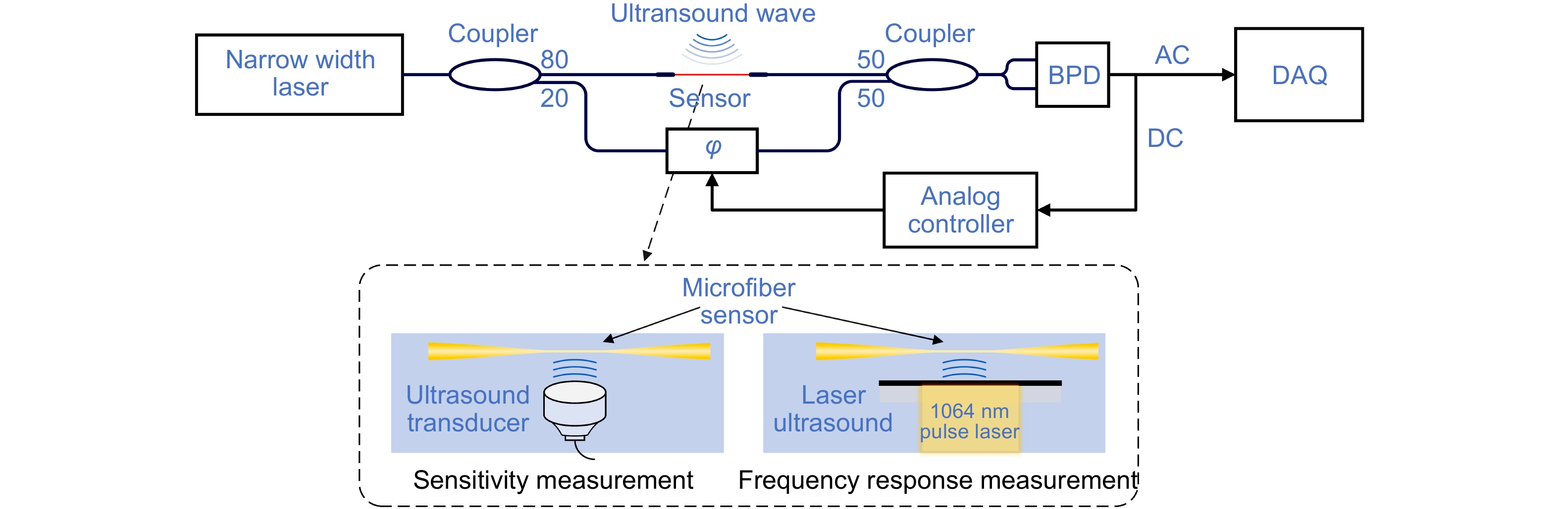
Figure 2.
The diagram of ultrasound detection system. φ: Phase modulator. BPD: Balanced photodetector. DC: Direct current signal. AC: Alternating current signal. DAQ: Data acquisition card.
-
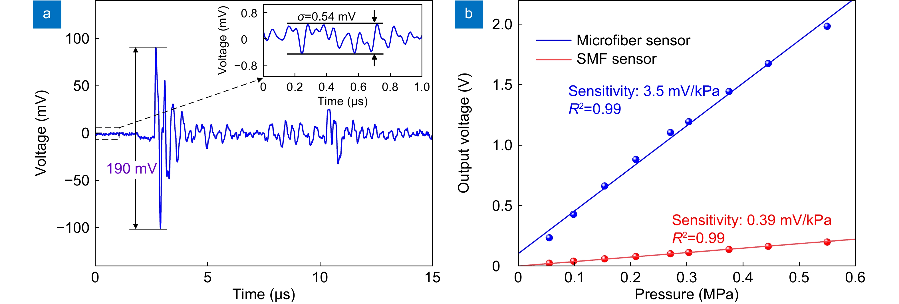
Figure 3.
(a) Time response of the sensor to an ultrasound pulse. (b) Ultrasound pressure sensitivities of microfiber sensor and SMF sensor.
-
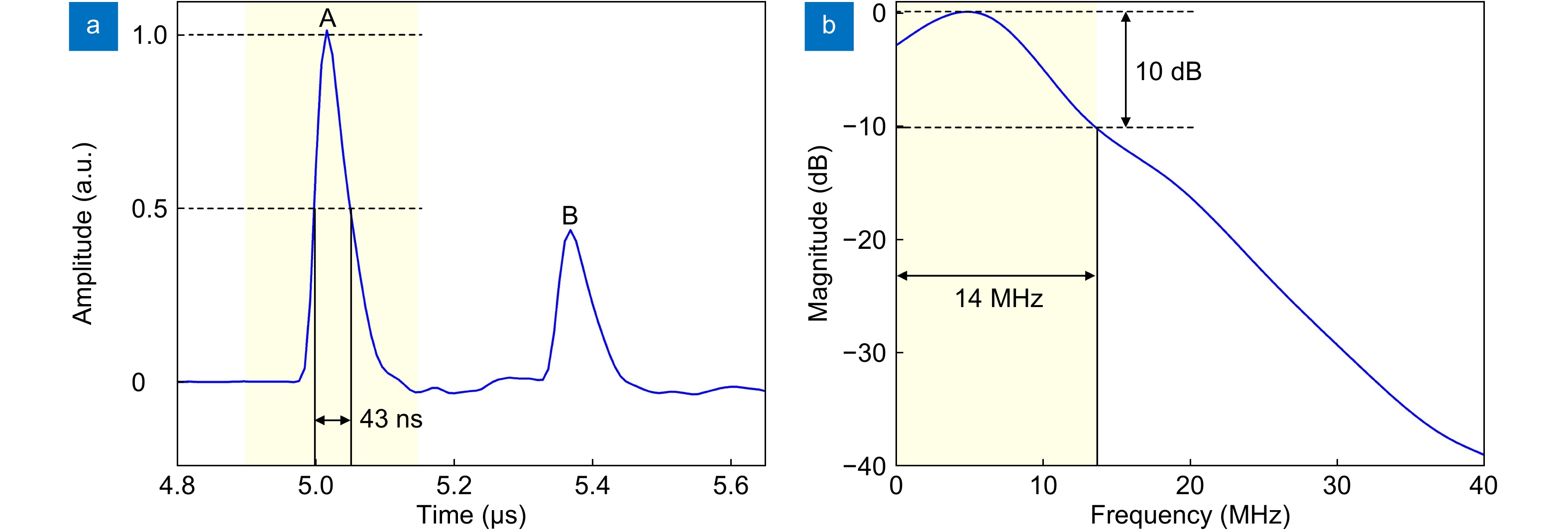
Figure 4.
(a) Recorded signals in response to broadband ultrasound wave. (b) Measured frequency responses of the microfiber-based ultrasound sensor.
-
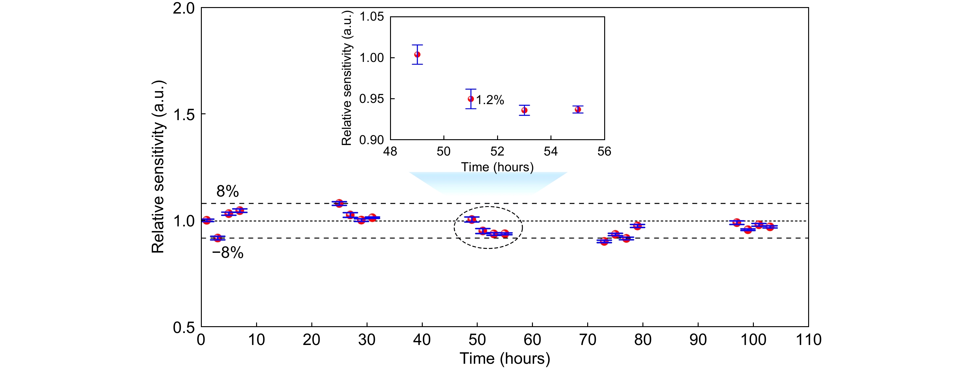
Figure 5.
The short-term and long-term stability of the microfiber-based ultrasound sensor.
-
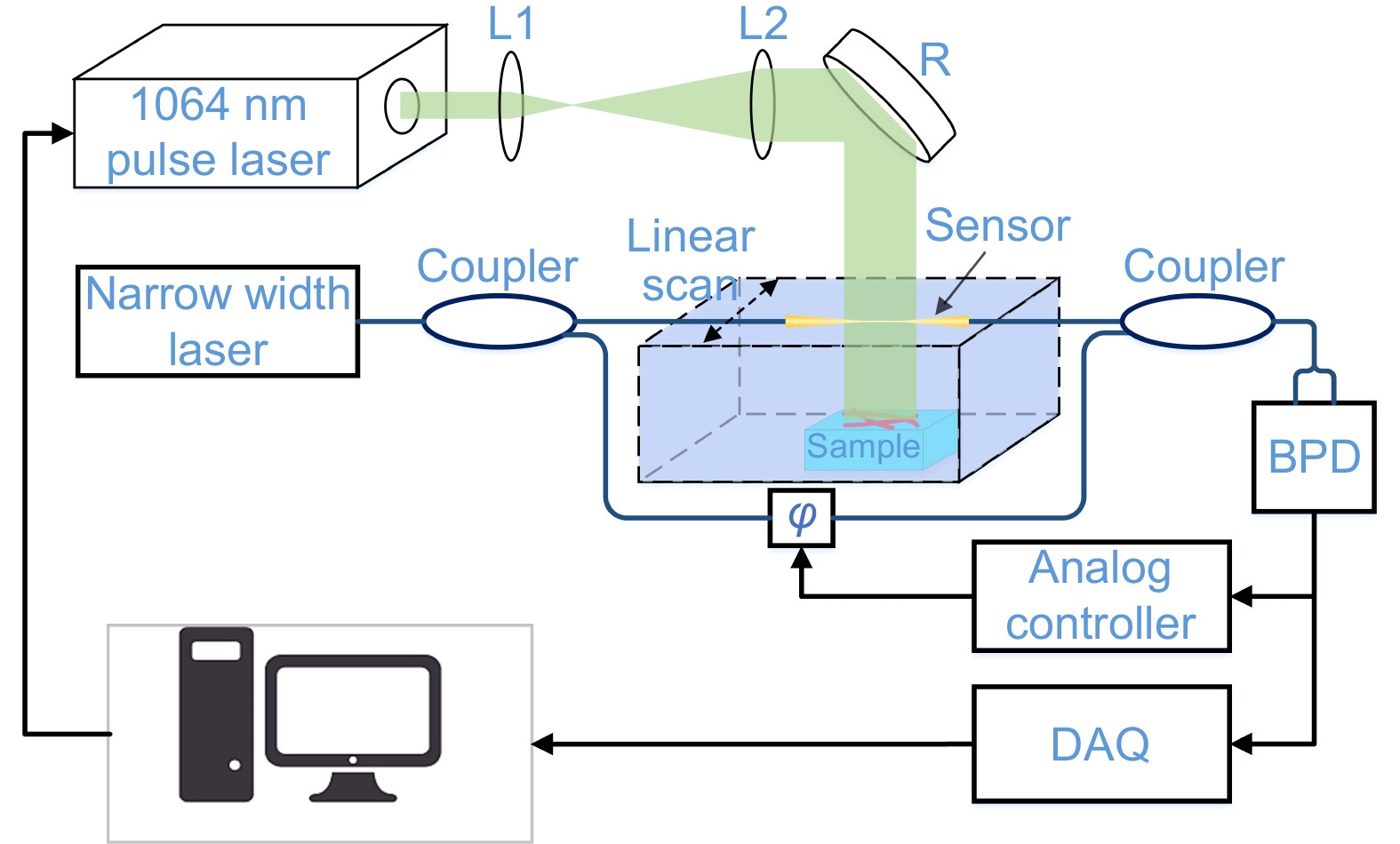
Figure 6.
Schematic of the photoacoustic tomography imaging system. L1&L2: Lens. R: Reflector.
-
Figure 7.
Images of three human hairs using the photoacoustic tomography system. (a, b) The sensor is placed perpendicular and parallel to hairs in the axial and lateral resolution measurements, respectively. (c, d) The reconstructed images of the hairs corresponding to (a) and (b), respectively. (e, f) The axial and lateral resolutions at different imaging depths.


