| Citation: | Jo Y, Park H, Yoon H et al. Advanced biological imaging techniques based on metasurfaces. Opto-Electron Adv 7, 240122 (2024). doi: 10.29026/oea.2024.240122 |
-
Abstract
Advanced imaging techniques have been widely used in various biological studies. Currently, numerous imaging modalities are utilized in biological applications, including medical imaging, diagnosis, biometrics, and fundamental biological research. Consequently, the demand for faster, clearer, and more accurate imaging techniques to support sophisticated biological studies has increased. However, there is a limitation in enhancing performance of imaging devices owing to the system complexity associated with bulky conventional optical elements. To address this issue, metasurfaces, which are flat and compact optical elements, have been considered potential candidates for biological imaging. Here, we comprehensively discuss the metasurface empowered various imaging applications in biology, including their working principles and design strategies. Furthermore, we compared conventional imaging modalities with the metasurface-based imaging system. Finally, we discuss the current challenges and offer future perspectives on metasurfaces.-
Keywords:
- metasurface /
- phase modulation /
- metamaterial /
- biological imaging techniques
-

-
References
[1] Avrutsky I, Chaganti K, Salakhutdinov I et al. Concept of a miniature optical spectrometer using integrated optical and micro-optical components. Appl Opt 45, 7811–7817 (2006). doi: 10.1364/AO.45.007811 [2] Khorasaninejad M, Capasso F. Metalenses: versatile multifunctional photonic components. Science 358, eaam8100 (2017). doi: 10.1126/science.aam8100 [3] Khorasaninejad M, Chen WT, Devlin RC et al. Metalenses at visible wavelengths: diffraction-limited focusing and subwavelength resolution imaging. Science 352, 1190–1194 (2016). doi: 10.1126/science.aaf6644 [4] Ni XJ, Ishii S, Kildishev AV et al. Ultra-thin, planar, Babinet-inverted plasmonic metalenses. Light Sci Appl 2, e72 (2013). doi: 10.1038/lsa.2013.28 [5] Kim I, Jang J, Kim G et al. Pixelated bifunctional metasurface-driven dynamic vectorial holographic color prints for photonic security platform. Nat Commun 12, 3614 (2021). doi: 10.1038/s41467-021-23814-5 [6] Kim I, Kim WS, Kim K et al. Holographic metasurface gas sensors for instantaneous visual alarms. Sci Adv 7, eabe9943 (2021). doi: 10.1126/sciadv.abe9943 [7] Paniagua-Dominguez R, Yu YF, Khaidarov E et al. A metalens with a near-unity numerical aperture. Nano Lett 18, 2124–2132 (2018). doi: 10.1021/acs.nanolett.8b00368 [8] Liang HW, Lin QL, Xie XS et al. Ultrahigh numerical aperture metalens at visible wavelengths. Nano Lett 18, 4460–4466 (2018). doi: 10.1021/acs.nanolett.8b01570 [9] Chung H, Miller OD. High-NA achromatic metalenses by inverse design. Opt Express 28, 6945–6965 (2020). doi: 10.1364/OE.385440 [10] Zhang SY, Wong CL, Zeng SW et al. Metasurfaces for biomedical applications: imaging and sensing from a nanophotonics perspective. Nanophotonics 10, 259–293 (2021). [11] Nguyen DD, Lee S, Kim I. Recent advances in metaphotonic biosensors. Biosensors 13, 631 (2023). doi: 10.3390/bios13060631 [12] Chen WT, Zhu AY, Sanjeev V et al. A broadband achromatic metalens for focusing and imaging in the visible. Nat Nanotechnol 13, 220–226 (2018). doi: 10.1038/s41565-017-0034-6 [13] Lin RJ, Su VC, Wang SM et al. Achromatic metalens array for full-colour light-field imaging. Nat Nanotechnol 14, 227–231 (2019). doi: 10.1038/s41565-018-0347-0 [14] Rho J, Ye ZL, Xiong Y et al. Spherical hyperlens for two-dimensional sub-diffractional imaging at visible frequencies. Nat Commun 1, 143 (2010). doi: 10.1038/ncomms1148 [15] Lee D, Kim YD, Kim M et al. Realization of wafer-scale hyperlens device for sub-diffractional biomolecular imaging. ACS Photonics 5, 2549–2554 (2018). doi: 10.1021/acsphotonics.7b01182 [16] Lee YU, Zhao JX, Ma Q et al. Metamaterial assisted illumination nanoscopy via random super-resolution speckles. Nat Commun 12, 1559 (2021). doi: 10.1038/s41467-021-21835-8 [17] Masuda S, Kuboki T, Kidoaki S et al. High axial and lateral resolutions on self-assembled gold nanoparticle metasurfaces for live-cell imaging. ACS Appl Nano Mater 3, 11135–11142 (2020). doi: 10.1021/acsanm.0c02300 [18] Huo PC, Zhang C, Zhu WQ et al. Photonic spin-multiplexing metasurface for switchable spiral phase contrast imaging. Nano Lett 20, 2791–2798 (2020). doi: 10.1021/acs.nanolett.0c00471 [19] Wang XW, Wang H, Wang JL et al. Single-shot isotropic differential interference contrast microscopy. Nat Commun 14, 2063 (2023). doi: 10.1038/s41467-023-37606-6 [20] Shi FH, Wen J, Lei DY. High-efficiency, large-area lattice light-sheet generation by dielectric metasurfaces. Nanophotonics 9, 4043–4051 (2020). doi: 10.1515/nanoph-2020-0227 [21] Luo Y, Tseng ML, Vyas S et al. Meta-lens light-sheet fluorescence microscopy for in vivo imaging. Nanophotonics 11, 1949–1959 (2022). doi: 10.1515/nanoph-2021-0748 [22] Wang CH, Chen QM, Liu HL et al. Miniature two-photon microscopic imaging using dielectric metalens. Nano Lett 23, 8256–8263 (2023). doi: 10.1021/acs.nanolett.3c02439 [23] Barulin A, Park H, Park B et al. Dual-wavelength UV-visible metalens for multispectral photoacoustic microscopy: a simulation study. Photoacoustics 32, 100545 (2023). doi: 10.1016/j.pacs.2023.100545 [24] Song W, Guo CK, Zhao YT et al. Ultraviolet metasurface-assisted photoacoustic microscopy with great enhancement in DOF for fast histology imaging. Photoacoustics 32, 100525 (2023). doi: 10.1016/j.pacs.2023.100525 [25] Pahlevaninezhad H, Khorasaninejad M, Huang YW et al. Nano-optic endoscope for high-resolution optical coherence tomography in vivo. Nat Photonics 12, 540–547 (2018). doi: 10.1038/s41566-018-0224-2 [26] Schmidt R, Slobozhanyuk A, Belov P et al. Flexible and compact hybrid metasurfaces for enhanced ultra high field in vivo magnetic resonance imaging. Sci Rep 7, 1678 (2017). doi: 10.1038/s41598-017-01932-9 [27] Lassalle E, Mass TWW, Eschimese D et al. Imaging properties of large field-of-view quadratic metalenses and their applications to fingerprint detection. ACS Photonics 8, 1457–1468 (2021). doi: 10.1021/acsphotonics.1c00237 [28] Hsu WC, Chang CH, Hong YH et al. Metasurface- and PCSEL-based structured light for monocular depth perception and facial recognition. Nano Lett 24, 1808–1815 (2024). doi: 10.1021/acs.nanolett.3c05002 [29] Yu NF, Genevet P, Kats MA et al. Light propagation with phase discontinuities: generalized laws of reflection and refraction. Science 334, 333–337 (2011). doi: 10.1126/science.1210713 [30] Hu J, Bandyopadhyay S, Liu YH et al. A review on metasurface: from principle to smart metadevices. Front Phys 8, 586087 (2021). doi: 10.3389/fphy.2020.586087 [31] Yu NF, Capasso F. Flat optics with designer metasurfaces. Nat Mater 13, 139–150 (2014). doi: 10.1038/nmat3839 [32] Khorasaninejad M, Zhu AY, Roques-Carmes C et al. Polarization-insensitive metalenses at visible wavelengths. Nano Lett 16, 7229–7234 (2016). doi: 10.1021/acs.nanolett.6b03626 [33] Khorasaninejad M, Chen WT, Oh J et al. Super-dispersive off-axis meta-lenses for compact high resolution spectroscopy. Nano Lett 16, 3732–3737 (2016). doi: 10.1021/acs.nanolett.6b01097 [34] Mueller JPB, Rubin NA, Devlin RC et al. Metasurface polarization optics: independent phase control of arbitrary orthogonal states of polarization. Phys Rev Lett 118, 113901 (2017). doi: 10.1103/PhysRevLett.118.113901 [35] Cong LQ, Xu NN, Zhang WL et al. Polarization control in terahertz metasurfaces with the lowest order rotational symmetry. Adv Opt Mater 3, 1176–1183 (2015). doi: 10.1002/adom.201500100 [36] Chen XZ, Huang LL, Mühlenbernd H et al. Dual-polarity plasmonic metalens for visible light. Nat Commun 3, 1198 (2012). doi: 10.1038/ncomms2207 [37] Arbabi A, Horie Y, Bagheri M et al. Dielectric metasurfaces for complete control of phase and polarization with subwavelength spatial resolution and high transmission. Nat Nanotechnol 10, 937–943 (2015). doi: 10.1038/nnano.2015.186 [38] Jiang SC, Xiong X, Hu YS et al. High-efficiency generation of circularly polarized light via symmetry-induced anomalous reflection. Phys Rev B 91, 125421 (2015). doi: 10.1103/PhysRevB.91.125421 [39] Huang LL, Chen XZ, Mühlenbernd H et al. Dispersionless phase discontinuities for controlling light propagation. Nano Lett 12, 5750–5755 (2012). doi: 10.1021/nl303031j [40] Moon SW, Lee C, Yang Y et al. Tutorial on metalenses for advanced flat optics: design, fabrication, and critical considerations. J Appl Phys 131, 091101 (2022). doi: 10.1063/5.0078804 [41] Luo WJ, Sun SL, Xu HX et al. Transmissive ultrathin pancharatnam-berry metasurfaces with nearly 100% efficiency. Phys Rev Appl 7, 044033 (2017). doi: 10.1103/PhysRevApplied.7.044033 [42] Fu X, Liang HW, Li JT. Metalenses: from design principles to functional applications. Front Optoelectron 14, 170–186 (2021). doi: 10.1007/s12200-021-1201-9 [43] Kim J, Li YM, Miskiewicz MN et al. Fabrication of ideal geometric-phase holograms with arbitrary wavefronts. Optica 2, 958–964 (2015). doi: 10.1364/OPTICA.2.000958 [44] Wen DD, Yue FY, Li GX et al. Helicity multiplexed broadband metasurface holograms. Nat Commun 6, 8241 (2015). doi: 10.1038/ncomms9241 [45] Chen WT, Khorasaninejad M, Zhu AY et al. Generation of wavelength-independent subwavelength Bessel beams using metasurfaces. Light Sci Appl 6, e16259 (2017). [46] Xu R, Chen P, Tang J et al. Perfect higher-order Poincaré sphere beams from digitalized geometric phases. Phys Rev Appl 10, 034061 (2018). doi: 10.1103/PhysRevApplied.10.034061 [47] Jeon D, Shin K, Moon SW et al. Recent advancements of metalenses for functional imaging. Nano Converg 10, 24 (2023). doi: 10.1186/s40580-023-00372-8 [48] Liu MK, Choi DY. Extreme Huygens’ metasurfaces based on quasi-bound states in the continuum. Nano Lett 18, 8062–8069 (2018). doi: 10.1021/acs.nanolett.8b04774 [49] Kim G, Kim Y, Yun J et al. Metasurface-driven full-space structured light for three-dimensional imaging. Nat Commun 13, 5920 (2022). doi: 10.1038/s41467-022-32117-2 [50] Wang YJ, Chen QM, Yang WH et al. High-efficiency broadband achromatic metalens for near-IR biological imaging window. Nat Commun 12, 5560 (2021). doi: 10.1038/s41467-021-25797-9 [51] Chen WT, Zhu AY, Sisler J et al. A broadband achromatic polarization-insensitive metalens consisting of anisotropic nanostructures. Nat Commun 10, 355 (2019). doi: 10.1038/s41467-019-08305-y [52] Heiden JT, Jang MS. Design framework for polarization-insensitive multifunctional achromatic metalenses. Nanophotonics 11, 583–591 (2022). doi: 10.1515/nanoph-2021-0638 [53] Faraji-Dana M, Arbabi E, Arbabi A et al. Compact folded metasurface spectrometer. Nat Commun 9, 4196 (2018). doi: 10.1038/s41467-018-06495-5 [54] Cai GY, Li YH, Zhang Y et al. Compact angle-resolved metasurface spectrometer. Nat Mater 23, 71–78 (2024). doi: 10.1038/s41563-023-01710-1 [55] Wang RX, Ansari MA, Ahmed H et al. Compact multi-foci metalens spectrometer. Light Sci Appl 12, 103 (2023). doi: 10.1038/s41377-023-01148-9 [56] Faraji-Dana M, Arbabi E, Arbabi A et al. Folded planar metasurface spectrometer. In Proceedings of 2018 Conference on Lasers and Electro-Optics (CLEO) 1–2 (IEEE, 2018). [57] Liu WQ, Deng L, Li SF et al. High transmittance and broadband group delay metasurface element in Ka band. In Proceedings of the 2021 IEEE 4th International Conference on Electronic Information and Communication Technology (ICEICT) 669–671 (IEEE, 2021); http://doi.org/10.1109/ICEICT53123.2021.9531170. [58] Miyamaru F, Morita H, Nishiyama Y et al. Ultrafast optical control of group delay of narrow-band terahertz waves. Sci Rep 4, 4346 (2014). doi: 10.1038/srep04346 [59] Jiang L, Li XZ, Wu QX et al. Neural network enabled metasurface design for phase manipulation. Opt Express 29, 2521–2528 (2021). doi: 10.1364/OE.413079 [60] Chen MZ, Cheng Q, Xia F et al. Metasurface‐based spatial phasers for analogue signal processing. Adv Opt Mater 8, 2000128 (2020). doi: 10.1002/adom.202000128 [61] Tsilipakos O, Koschny T, Soukoulis CM. Antimatched electromagnetic metasurfaces for broadband arbitrary phase manipulation in reflection. ACS Photonics 5, 1101–1107 (2018). doi: 10.1021/acsphotonics.7b01415 [62] Trubetskov MK, Von Pechmann M, Angelov IB et al. Measurements of the group delay and the group delay dispersion with resonance scanning interferometer. Opt Express 21, 6658–6669 (2013). doi: 10.1364/OE.21.006658 [63] Song NT, Xu NX, Shan DZ et al. Broadband achromatic metasurfaces for longwave infrared applications. Nanomaterials (Basel) 11 , 2760 (2021). [64] He YL, Song BX, Tang J. Optical metalenses: fundamentals, dispersion manipulation, and applications. Front Optoelectron 15, 24 (2022). doi: 10.1007/s12200-022-00017-4 [65] Pan Y, Neuss S, Leifert A et al. Size‐dependent cytotoxicity of gold nanoparticles. Small 3, 1941–1949 (2007). doi: 10.1002/smll.200700378 [66] Gonçalves MR, Minassian H, Melikyan A. Plasmonic resonators: fundamental properties and applications. J Phys D Appl Phys 53, 443002 (2020). doi: 10.1088/1361-6463/ab96e9 [67] Xu Q, Zhang XQ, Xu YH et al. Plasmonic metalens based on coupled resonators for focusing of surface plasmons. Sci Rep 6, 37861 (2016). doi: 10.1038/srep37861 [68] Autore M, Hillenbrand R. What momentum mismatch. Nat Nanotechnol 14, 308–309 (2019). doi: 10.1038/s41565-019-0390-5 [69] Kim K, Oh Y, Ma K et al. Plasmon-enhanced total-internal-reflection fluorescence by momentum-mismatched surface nanostructures. Opt Lett 34, 3905–3907 (2009). doi: 10.1364/OL.34.003905 [70] Qin LS, Chen X, Zhang LL et al. Design, fabrication and testing of gain SPR sensor chip. J Phys Conf Ser 1209, 012006 (2019). doi: 10.1088/1742-6596/1209/1/012006 [71] Petryayeva E, Krull UJ. Localized surface plasmon resonance: nanostructures, bioassays and biosensing—A review. Anal Chim Acta 706, 8–24 (2011). doi: 10.1016/j.aca.2011.08.020 [72] Palermo G, Lininger A, Guglielmelli A et al. All-optical tunability of metalenses permeated with liquid crystals. ACS nano 16, 16539–16548 (2022). doi: 10.1021/acsnano.2c05887 [73] Park S, Hahn JW, Lee JY. Doubly resonant metallic nanostructure for high conversion efficiency of second harmonic generation. Opt Express 20, 4856–4870 (2012). doi: 10.1364/OE.20.004856 [74] Damgaard-Carstensen C, Ding F, Meng C et al. Demonstration of> 2π reflection phase range in optical metasurfaces based on detuned gap-surface plasmon resonators. Sci Rep 10, 19031 (2020). doi: 10.1038/s41598-020-75931-8 [75] Yao K, Liu YM. Plasmonic metamaterials. Nanotechnol Rev 3, 177–210 (2014). [76] Tong XC. Plasmonic metamaterials and metasurfaces. In Tong XC. Functional Metamaterials and Metadevices 129–153 (Springer, Cham, 2018). [77] Christopher P, Xin HL, Linic S. Visible-light-enhanced catalytic oxidation reactions on plasmonic silver nanostructures. Nat Chem 3, 467–472 (2011). doi: 10.1038/nchem.1032 [78] Barnard ES, White JS, Chandran A et al. Spectral properties of plasmonic resonator antennas. Opt Express 16, 16529–16537 (2008). doi: 10.1364/OE.16.016529 [79] Sun SL, He Q, Hao JM et al. Electromagnetic metasurfaces: physics and applications. Adv Opt Photonics 11, 380–479 (2019). doi: 10.1364/AOP.11.000380 [80] Pors A, Albrektsen O, Radko IP et al. Gap plasmon-based metasurfaces for total control of reflected light. Sci Rep 3, 2155 (2013). doi: 10.1038/srep02155 [81] Ding F, Yang YQ, Deshpande RA et al. A review of gap-surface plasmon metasurfaces: fundamentals and applications. Nanophotonics 7, 1129–1156 (2018). doi: 10.1515/nanoph-2017-0125 [82] Pors A, Bozhevolnyi SI. Plasmonic metasurfaces for efficient phase control in reflection. Opt Express 21, 27438–27451 (2013). doi: 10.1364/OE.21.027438 [83] Pors A, Nielsen MG, Eriksen RL et al. Broadband focusing flat mirrors based on plasmonic gradient metasurfaces. Nano Lett 13, 829–834 (2013). doi: 10.1021/nl304761m [84] Boroviks S, Deshpande RA, Mortensen NA et al. Multifunctional metamirror: polarization splitting and focusing. ACS Photonics 5, 1648–1653 (2018). doi: 10.1021/acsphotonics.7b01091 [85] Cortés E, Wendisch FJ, Sortino L et al. Optical metasurfaces for energy conversion. Chem Rev 122, 15082–15176 (2022). doi: 10.1021/acs.chemrev.2c00078 [86] Linic S, Christopher P, Ingram DB. Plasmonic-metal nanostructures for efficient conversion of solar to chemical energy. Nat Mater 10, 911–921 (2011). doi: 10.1038/nmat3151 [87] Gutruf P, Zou CJ, Withayachumnankul W et al. Mechanically tunable dielectric resonator metasurfaces at visible frequencies. Acs Nano 10, 133–141 (2016). doi: 10.1021/acsnano.5b05954 [88] Campione S, Basilio LI, Warne LK et al. Tailoring dielectric resonator geometries for directional scattering and Huygens’ metasurfaces. Opt Express 23, 2293–2307 (2015). doi: 10.1364/OE.23.002293 [89] Decker M, Staude I, Falkner M et al. High‐efficiency dielectric Huygens’ surfaces. Adv Opt Mater 3, 813–820 (2015). doi: 10.1002/adom.201400584 [90] Maier SA. Plasmonics: Fundamentals and Applications (Springer, New York, 2007). [91] Koshelev K, Kivshar Y. Dielectric resonant metaphotonics. ACS Photonics 8, 102–112 (2021). [92] Lin DM, Fan PY, Hasman E et al. Dielectric gradient metasurface optical elements. Science 345, 298–302 (2014). doi: 10.1126/science.1253213 [93] Devlin RC, Khorasaninejad M, Chen WT et al. Broadband high-efficiency dielectric metasurfaces for the visible spectrum. Proc Natl Acad Sci USA 113, 10473–10478 (2016). doi: 10.1073/pnas.1611740113 [94] Zhou SH, Shen ZX, Li XA et al. Liquid crystal integrated metalens with dynamic focusing property. Opt Lett 45, 4324–4327 (2020). doi: 10.1364/OL.398601 [95] Pan MY, Fu YF, Zheng MJ et al. Dielectric metalens for miniaturized imaging systems: progress and challenges. Light Sci Appl 11, 195 (2022). doi: 10.1038/s41377-022-00885-7 [96] Kerker M, Wang DS, Giles CL. Electromagnetic scattering by magnetic spheres. J Opt Soc Am 73, 765–767 (1983). doi: 10.1364/JOSA.73.000765 [97] Bukharin MM, Pecherkin VY, Ospanova AK et al. Transverse Kerker effect in all-dielectric spheroidal particles. Sci Rep 12, 7997 (2022). doi: 10.1038/s41598-022-11733-4 [98] Liu W, Kivshar YS. Generalized Kerker effects in nanophotonics and meta-optics [Invited]. Opt Express 26, 13085–13105 (2018). doi: 10.1364/OE.26.013085 [99] Liu WW, Li ZC, Cheng H et al. Dielectric resonance-based optical metasurfaces: from fundamentals to applications. iScience 23, 101868 (2020). doi: 10.1016/j.isci.2020.101868 [100] Genevet P, Capasso F, Aieta F et al. Recent advances in planar optics: from plasmonic to dielectric metasurfaces. Optica 4, 139–152 (2017). doi: 10.1364/OPTICA.4.000139 [101] Long S, McAllister M, Shen L. The resonant cylindrical dielectric cavity antenna. IEEE Trans Antennas Propag 31, 406–412 (1983). doi: 10.1109/TAP.1983.1143080 [102] Wang SW, Lai JJ, Wu T et al. Wide-band achromatic metalens for visible light by dispersion compensation method. J Phys D Appl Phys 50, 455101 (2017). doi: 10.1088/1361-6463/aa8c90 [103] Rytov S. Electromagnetic properties of a finely stratified medium. Sov Phys JEPT 2, 466–475 (1956). [104] Vo S, Fattal D, Sorin WV et al. Sub-wavelength grating lenses with a twist. IEEE Photonics Technol Lett 26, 1375–1378 (2014). doi: 10.1109/LPT.2014.2325947 [105] Arbabi A, Horie Y, Ball AJ et al. Subwavelength-thick lenses with high numerical apertures and large efficiency based on high-contrast transmitarrays. Nat Commun 6, 7069 (2015). doi: 10.1038/ncomms8069 [106] Powell DA. Interference between the modes of an all-dielectric meta-atom. Phys Rev Appl 7, 034006 (2017). doi: 10.1103/PhysRevApplied.7.034006 [107] Wang Q, Rogers ETF, Gholipour B et al. Optically reconfigurable metasurfaces and photonic devices based on phase change materials. Nat Photonics 10, 60–65 (2016). doi: 10.1038/nphoton.2015.247 [108] Zhang YF, Fowler C, Liang JH et al. Electrically reconfigurable non-volatile metasurface using low-loss optical phase-change material. Nat Nanotechnol 16, 661–666 (2021). doi: 10.1038/s41565-021-00881-9 [109] Abdollahramezani S, Hemmatyar O, Taghinejad M et al. Electrically driven reprogrammable phase-change metasurface reaching 80% efficiency. Nat Commun 13, 1696 (2022). doi: 10.1038/s41467-022-29374-6 [110] Wang Y, Cui DJ, Wang Y et al. Electrically and thermally tunable multifunctional terahertz metasurface array. Phys Rev A 105, 033520 (2022). doi: 10.1103/PhysRevA.105.033520 [111] Shao ZW, Cao X, Luo HJ et al. Recent progress in the phase-transition mechanism and modulation of vanadium dioxide materials. NPG Asia Mater 10, 581–605 (2018). doi: 10.1038/s41427-018-0061-2 [112] Shalaginov MY, An SS, Zhang YF et al. Reconfigurable all-dielectric metalens with diffraction-limited performance. Nat Commun 12, 1225 (2021). doi: 10.1038/s41467-021-21440-9 [113] Zhang WH, Wu XQ, Li L et al. Fabrication of a VO2-based tunable metasurface by electric-field scanning probe lithography with precise depth control. ACS Appl Mater Interfaces 15, 13517–13525 (2023). doi: 10.1021/acsami.2c21935 [114] Hashemi MRM, Yang SH, Wang TY et al. Electronically-controlled beam-steering through vanadium dioxide metasurfaces. Sci Rep 6, 35439 (2016). doi: 10.1038/srep35439 [115] King J, Wan CH, Park TJ et al. Electrically tunable VO2–metal metasurface for mid-infrared switching, limiting and nonlinear isolation. Nat Photonics 18, 74–80 (2024). doi: 10.1038/s41566-023-01324-8 [116] Wan CH, Zhang Z, Woolf D et al. On the optical properties of thin‐film vanadium dioxide from the visible to the far infrared. Ann Phys 531, 1900188 (2019). doi: 10.1002/andp.201900188 [117] Chu F, Tian LL, Li R et al. Adaptive nematic liquid crystal lens array with resistive layer. Liq Cryst 47, 563–571 (2020). doi: 10.1080/02678292.2019.1662502 [118] Kang DH, Heo HS, Yang YH et al. Liquid crystal-integrated metasurfaces for an active photonic platform. Opto-Electron Adv 7, 230216 (2024). doi: 10.29026/oea.2024.230216 [119] Chang X, Pivnenko M, Shrestha P et al. Electrically tuned active metasurface towards metasurface-integrated liquid crystal on silicon (meta-LCoS) devices. Opt Express 31, 5378–5387 (2023). doi: 10.1364/OE.483452 [120] Ji YY, Fan F, Zhang X et al. Active terahertz anisotropy and dispersion engineering based on dual-frequency liquid crystal and dielectric metasurface. J Lightwave Technol 38, 4030–4036 (2020). [121] Rocco D, Carletti L, Caputo R et al. Switching the second harmonic generation by a dielectric metasurface via tunable liquid crystal. Opt Express 28, 12037–12046 (2020). doi: 10.1364/OE.386776 [122] Badloe T, Kim Y, Kim J et al. Bright-field and edge-enhanced imaging using an electrically tunable dual-mode metalens. ACS Nano 17, 14678–14685 (2023). doi: 10.1021/acsnano.3c02471 [123] Bosch M, Shcherbakov MR, Won K et al. Electrically actuated varifocal lens based on liquid-crystal-embedded dielectric metasurfaces. Nano Lett 21, 3849–3856 (2021). doi: 10.1021/acs.nanolett.1c00356 [124] Kamali SM, Arbabi A, Arbabi E et al. Decoupling optical function and geometrical form using conformal flexible dielectric metasurfaces. Nat Commun 7, 11618 (2016). doi: 10.1038/ncomms11618 [125] Kamali SM, Arbabi E, Arbabi A et al. Highly tunable elastic dielectric metasurface lenses. Laser Photonics Rev 10, 1002–1008 (2016). doi: 10.1002/lpor.201600144 [126] Li J, Fan HJ, Ye H et al. Design of multifunctional tunable metasurface assisted by elastic substrate. Nanomaterials 12, 2387 (2022). doi: 10.3390/nano12142387 [127] Li WL, He P, Lei DY et al. Super-resolution multicolor fluorescence microscopy enabled by an apochromatic super-oscillatory lens with extended depth-of-focus. Nat Commun 14, 5107 (2023). doi: 10.1038/s41467-023-40725-9 [128] Ren YX, He HS, Tang HJ et al. Non-diffracting light wave: fundamentals and biomedical applications. Front Phys 9, 698343 (2021). doi: 10.3389/fphy.2021.698343 [129] Bouchal Z. Nondiffracting optical beams: physical properties, experiments, and applications. Czech J Phys 53, 537–578 (2003). doi: 10.1023/A:1024802801048 [130] Li DC, Wang XR, Ling JZ et al. Planar efficient metasurface for generation of Bessel beam and super-resolution focusing. Opt Quant Electron 53, 143 (2021). doi: 10.1007/s11082-021-02765-7 [131] Lin ZM, Li XW, Zhao RZ et al. High-efficiency Bessel beam array generation by Huygens metasurfaces. Nanophotonics 8, 1079–1085 (2019). doi: 10.1515/nanoph-2019-0085 [132] Shi FH, Qiu M, Zhang L et al. Multiplane illumination enabled by Fourier-transform metasurfaces for high-speed light-sheet microscopy. ACS Photonics 5, 1676–1684 (2018). doi: 10.1021/acsphotonics.7b01457 [133] Li CS, Guo YH, Chang XZ et al. A metasurface-on-fiber light-sheet generator for biological imaging. Opt Commun 559, 130378 (2024). doi: 10.1016/j.optcom.2024.130378 [134] Luo Y, Tseng ML, Vyas S et al. Metasurface‐based abrupt autofocusing beam for biomedical applications. Small Methods 6, 2101228 (2022). doi: 10.1002/smtd.202101228 [135] Lu RW, Liang YJ, Meng GH et al. Rapid mesoscale volumetric imaging of neural activity with synaptic resolution. Nat Methods 17, 291–294 (2020). doi: 10.1038/s41592-020-0760-9 [136] Wang XW, Nie ZQ, Liang Y et al. Recent advances on optical vortex generation. Nanophotonics 7, 1533–1556 (2018). doi: 10.1515/nanoph-2018-0072 [137] Wang S, Li L, Wen S et al. Metalens for accelerated optoelectronic edge detection under ambient illumination. Nano Lett 24, 356–361 (2023). [138] Zheludev NI. What diffraction limit. Nat Mater 7, 420–422 (2008). doi: 10.1038/nmat2163 [139] Hänninen P. Beyond the diffraction limit. Nature 419, 802 (2002). [140] Park SC, Park MK, Kang MG. Super-resolution image reconstruction: a technical overview. IEEE Signal Process Mag 20, 21–36 (2003). doi: 10.1109/MSP.2003.1203207 [141] Huang B, Babcock H, Zhuang XW. Breaking the diffraction barrier: super-resolution imaging of cells. Cell 143, 1047–1058 (2010). doi: 10.1016/j.cell.2010.12.002 [142] Bliokh KY, Bekshaev AY, Nori F. Extraordinary momentum and spin in evanescent waves. Nat Commun 5, 3300 (2014). doi: 10.1038/ncomms4300 [143] Loerke D, Preitz B, Stuhmer W et al. Super-resolution measurements with evanescent-wave fluorescence-excitation using variable beam incidence. J Biomed Opt 5, 23–30 (2000). doi: 10.1117/1.429964 [144] Pendry JB. Negative refraction makes a perfect lens. Phys Rev Lett 85, 3966–3969 (2000). doi: 10.1103/PhysRevLett.85.3966 [145] Fang N, Lee H, Sun C et al. Sub-diffraction-limited optical imaging with a silver superlens. Science 308, 534–537 (2005). doi: 10.1126/science.1108759 [146] Lu D, Liu ZW. Hyperlenses and metalenses for far-field super-resolution imaging. Nat Commun 3, 1205 (2012). doi: 10.1038/ncomms2176 [147] Kim M, So S, Yao K et al. Deep sub-wavelength nanofocusing of UV-visible light by hyperbolic metamaterials. Sci Rep 6, 38645 (2016). doi: 10.1038/srep38645 [148] Jacob Z, Alekseyev LV, Narimanov E. Optical hyperlens: far-field imaging beyond the diffraction limit. Opt Express 14, 8247–8256 (2006). doi: 10.1364/OE.14.008247 [149] Liu ZW, Lee H, Xiong Y et al. Far-field optical hyperlens magnifying sub-diffraction-limited objects. Science 315, 1686 (2007). doi: 10.1126/science.1137368 [150] Kastrup L, Blom H, Eggeling C et al. Fluorescence fluctuation spectroscopy in subdiffraction focal volumes. Phys Rev Lett 94, 178104 (2005). doi: 10.1103/PhysRevLett.94.178104 [151] Honigmann A, Mueller V, Ta H et al. Scanning STED-FCS reveals spatiotemporal heterogeneity of lipid interaction in the plasma membrane of living cells. Nat Commun 5, 5412 (2014). doi: 10.1038/ncomms6412 [152] Barulin A, Kim I. Hyperlens for capturing sub-diffraction nanoscale single molecule dynamics. Opt Express 31, 12162–12174 (2023). doi: 10.1364/OE.486702 [153] Wei FF, Ponsetto JL, Liu ZW. Plasmonic structured illumination microscopy. In Liu ZW. Plasmonics and Super-Resolution Imaging 127–163 (Jenny Stanford Publishing, New York, 2017). [154] Wei FF, Lu D, Shen H et al. Wide field super-resolution surface imaging through plasmonic structured illumination microscopy. Nano Lett 14, 4634–4639 (2014). doi: 10.1021/nl501695c [155] Wei SB, Lei T, Du LP et al. Sub-100nm resolution PSIM by utilizing modified optical vortices with fractional topological charges for precise phase shifting. Opt Express 23, 30143–30148 (2015). doi: 10.1364/OE.23.030143 [156] Ponsetto JL, Wei FF, Liu ZW. Localized plasmon assisted structured illumination microscopy for wide-field high-speed dispersion-independent super resolution imaging. Nanoscale 6, 5807–5812 (2014). doi: 10.1039/C4NR00443D [157] Ponsetto JL, Bezryadina A, Wei FF et al. Experimental demonstration of localized plasmonic structured illumination microscopy. ACS Nano 11, 5344–5350 (2017). doi: 10.1021/acsnano.7b01158 [158] Lee YU, Li SL, Wisna GBM et al. Hyperbolic material enhanced scattering nanoscopy for label-free super-resolution imaging. Nat Commun 13, 6631 (2022). doi: 10.1038/s41467-022-34553-6 [159] Rogers ETF, Lindberg J, Roy T et al. A super-oscillatory lens optical microscope for subwavelength imaging. Nat Mater 11, 432–435 (2012). doi: 10.1038/nmat3280 [160] Berry MV, Popescu S. Evolution of quantum superoscillations and optical superresolution without evanescent waves. J Phys A: Math Gen 39, 6965–6977 (2006). doi: 10.1088/0305-4470/39/22/011 [161] Aharonov Y, Albert DZ, Vaidman L. How the result of a measurement of a component of the spin of a spin-1/2 particle can turn out to be 100. Phys Rev Lett 60, 1351–1354 (1988). doi: 10.1103/PhysRevLett.60.1351 [162] Huang FM, Zheludev N, Chen YF et al. Focusing of light by a nanohole array. Appl Phys Lett 90, 091119 (2007). doi: 10.1063/1.2710775 [163] Zheludev NI, Yuan G. Optical superoscillation technologies beyond the diffraction limit. Nat Rev Phys 4, 16–32 (2022). [164] Chen G, Wen ZQ, Qiu CW. Superoscillation: from physics to optical applications. Light Sci Appl 8, 56 (2019). doi: 10.1038/s41377-019-0163-9 [165] Qin F, Huang K, Wu JF et al. A supercritical lens optical label‐free microscopy: sub‐diffraction resolution and ultra‐long working distance. Adv Mater 29, 1602721 (2017). doi: 10.1002/adma.201602721 [166] Li Z, Zhang T, Wang YQ et al. Achromatic Broadband super‐resolution imaging by super‐oscillatory metasurface. Laser Photonics Rev 12, 1800064 (2018). doi: 10.1002/lpor.201800064 [167] Yuan GH, Rogers ETF, Zheludev NI. Achromatic super-oscillatory lenses with sub-wavelength focusing. Light Sci Appl 6, e17036 (2017). doi: 10.1038/lsa.2017.36 [168] Rogers ETF, Savo S, Lindberg J et al. Super-oscillatory optical needle. Appl Phys Lett 102, 031108 (2013). doi: 10.1063/1.4774385 [169] Roy T, Rogers ETF, Yuan GH et al. Point spread function of the optical needle super-oscillatory lens. Appl Phys Lett 104, 231109 (2014). doi: 10.1063/1.4882246 [170] Yuan GH, Rogers ETF, Roy T et al. Planar super-oscillatory lens for sub-diffraction optical needles at violet wavelengths. Sci Rep 4, 6333 (2014). doi: 10.1038/srep06333 [171] Diao JS, Yuan WZ, Yu YT et al. Controllable design of super-oscillatory planar lenses for sub-diffraction-limit optical needles. Opt Express 24, 1924–1933 (2016). doi: 10.1364/OE.24.001924 [172] Chen G, Wu ZX, Yu AP et al. Planar binary-phase lens for super-oscillatory optical hollow needles. Sci Rep 7, 4697 (2017). doi: 10.1038/s41598-017-05060-2 [173] Solli DR, Jalali B. Analog optical computing. Nat Photonics 9, 704–706 (2015). doi: 10.1038/nphoton.2015.208 [174] He SS, Wang RS, Luo HL. Computing metasurfaces for all-optical image processing: a brief review. Nanophotonics 11, 1083–1108 (2022). doi: 10.1515/nanoph-2021-0823 [175] Selinummi J, Ruusuvuori P, Podolsky I et al. Bright field microscopy as an alternative to whole cell fluorescence in automated analysis of macrophage images. PLoS One 4, e7497 (2009). doi: 10.1371/journal.pone.0007497 [176] Marr D, Hildreth E. Theory of edge detection. Proc Roy Soc B Biol Sci 207, 187–217 (1980). [177] Canny J. A computational approach to edge detection. IEEE Trans Pattern Anal Mach Intell PAMI-8, 679–698 (1986). doi: 10.1109/TPAMI.1986.4767851 [178] Maini R, Aggarwal H. Study and comparison of various image edge detection techniques. Int J Image Process 3, 1–12 (2009). doi: 10.1049/iet-ipr:20080080 [179] Wang JK, Zhang WH, Qi QQ et al. Gradual edge enhancement in spiral phase contrast imaging with fractional vortex filters. Sci Rep 5, 15826 (2015). doi: 10.1038/srep15826 [180] Wei SB, Zhu SW, Yuan XC. Image edge enhancement in optical microscopy with a Bessel-like amplitude modulated spiral phase filter. J Opt 13, 105704 (2011). doi: 10.1088/2040-8978/13/10/105704 [181] Yuan S, Xiang D, Liu XM et al. Edge detection based on computational ghost imaging with structured illuminations. Opt Commun 410, 350–355 (2018). doi: 10.1016/j.optcom.2017.10.016 [182] Zhou Y, Zheng HY, Kravchenko II et al. Flat optics for image differentiation. Nat Photonics 14, 316–323 (2020). doi: 10.1038/s41566-020-0591-3 [183] Chen MK, Wu YF, Feng L et al. Principles, functions, and applications of optical meta‐lens. Adv Opt Mater 9, 2001414 (2021). doi: 10.1002/adom.202001414 [184] Abdollahramezani S, Hemmatyar O, Adibi A. Meta-optics for spatial optical analog computing. Nanophotonics 9, 4075–4095 (2020). doi: 10.1515/nanoph-2020-0285 [185] Wan L, Pan DP, Yang SF et al. Optical analog computing of spatial differentiation and edge detection with dielectric metasurfaces. Opt Lett 45, 2070–2073 (2020). doi: 10.1364/OL.386986 [186] Zhou JX, Qian HL, Zhao JX et al. Two-dimensional optical spatial differentiation and high-contrast imaging. Natl Sci Rev 8, nwaa176 (2021). doi: 10.1093/nsr/nwaa176 [187] Guo C, Xiao M, Minkov M et al. Photonic crystal slab Laplace operator for image differentiation. Optica 5, 251–256 (2018). doi: 10.1364/OPTICA.5.000251 [188] Kim Y, Lee GY, Sung J et al. Spiral metalens for phase contrast imaging. Adv Funct Mater 32, 2106050 (2022). doi: 10.1002/adfm.202106050 [189] Dong HG, Wang FQ, Liang RS et al. Visible-wavelength metalenses for diffraction-limited focusing of double polarization and vortex beams. Opt Mater Express 7, 4029–4037 (2017). doi: 10.1364/OME.7.004029 [190] Zhou C, Chen YJ, Li Y et al. Laplace differentiator based on metasurface with toroidal dipole resonance. Adv Funct Mater 34, 2313777 (2024). doi: 10.1002/adfm.202313777 [191] Chu CH, Chia YH, Hsu HC et al. Intelligent phase contrast meta-microscope system. Nano Lett 23, 11630–11637 (2023). doi: 10.1021/acs.nanolett.3c03484 [192] Overvig A, Alù A. Diffractive nonlocal metasurfaces. Laser Photonics Rev 16, 2100633 (2022). doi: 10.1002/lpor.202100633 [193] Shastri K, Monticone F. Nonlocal flat optics. Nat Photonics 17, 36–47 (2023). doi: 10.1038/s41566-022-01098-5 [194] Kim M, Lee D, Kim J et al. Nonlocal metasurfaces‐enabled analog light localization for imaging and lithography. Laser Photonics Rev 18, 2300718 (2024). doi: 10.1002/lpor.202300718 [195] Li, Shiyu, and Chia Wei Hsu. Thickness bound for nonlocal wide-field-of-view metalenses. Light: Science & Applications 11.1, 338 (2022). doi: 10.1103/PhysRevLett.121.173004 [196] Kwon H, Sounas D, Cordaro A et al. Nonlocal metasurfaces for optical signal processing. Phys Rev Lett 121 , 173004 (2018). [197] Kwon H, Cordaro A, Sounas D et al. Dual-polarization analog 2D image processing with nonlocal metasurfaces. ACS Photonics 7, 1799–1805 (2020). doi: 10.1021/acsphotonics.0c00473 [198] Arnison MR, Larkin KG, Sheppard CJR et al. Linear phase imaging using differential interference contrast microscopy. J Microsc 214, 7–12 (2004). doi: 10.1111/j.0022-2720.2004.01293.x [199] Zernike F. How I discovered phase contrast. Science 121, 345–349 (1955). doi: 10.1126/science.121.3141.345 [200] Preza C, Snyder DL, Conchello JA. Theoretical development and experimental evaluation of imaging models for differential-interference-contrast microscopy. J Opt Soc Am A 16, 2185–2199 (1999). doi: 10.1364/JOSAA.16.002185 [201] Preza C. Rotational-diversity phase estimation from differential-interference-contrast microscopy images. J Opt Soc Am A 17, 415–424 (2000). doi: 10.1364/JOSAA.17.000415 [202] Shribak M. Quantitative orientation-independent differential interference contrast microscope with fast switching shear direction and bias modulation. J Opt Soc Am A 30, 769–782 (2013). doi: 10.1364/JOSAA.30.000769 [203] Zhao Q, Kang L, Du B et al. Electrically tunable negative permeability metamaterials based on nematic liquid crystals. Appl Phys Lett 90, 011112 (2007). doi: 10.1063/1.2430485 [204] Kang B, Woo JH, Choi E et al. Optical switching of near infrared light transmission in metamaterial-liquid crystal cell structure. Opt Express 18, 16492–16498 (2010). doi: 10.1364/OE.18.016492 [205] Zhang FL, Zhang WH, Zhao Q et al. Electrically controllable fishnet metamaterial based on nematic liquid crystal. Opt Express 19, 1563–1568 (2011). doi: 10.1364/OE.19.001563 [206] Su H, Wang H, Zhao H et al. Liquid-crystal-based electrically tuned electromagnetically induced transparency metasurface switch. Sci Rep 7, 17378 (2017). doi: 10.1038/s41598-017-17612-7 [207] Yu P, Li JX, Liu N. Electrically tunable optical metasurfaces for dynamic polarization conversion. Nano Lett 21, 6690–6695 (2021). doi: 10.1021/acs.nanolett.1c02318 [208] Badloe T, Kim I, Kim Y et al. Electrically tunable bifocal metalens with diffraction‐limited focusing and imaging at visible wavelengths. Adv Sci 8, 2102646 (2021). doi: 10.1002/advs.202102646 [209] Zhou SL, Wu YF, Chen SR et al. Phase change induced active metasurface devices for dynamic wavefront control. J Phys D Appl Phys 53, 204001 (2020). doi: 10.1088/1361-6463/ab7560 [210] Ee HS, Agarwal R. Tunable metasurface and flat optical zoom lens on a stretchable substrate. Nano Lett 16, 2818–2823 (2016). doi: 10.1021/acs.nanolett.6b00618 [211] Tseng ML, Hsiao HH, Chu CH et al. Metalenses: advances and applications. Adv Opt Mater 6, 1800554 (2018). doi: 10.1002/adom.201800554 [212] Dirdal CA, Thrane PCV, Dullo FT et al. MEMS-tunable dielectric metasurface lens using thin-film PZT for large displacements at low voltages. Opt Lett 47, 1049–1052 (2022). doi: 10.1364/OL.451750 [213] Han ZY, Colburn S, Majumdar A et al. MEMS-actuated metasurface Alvarez lens. Microsystems 6, 79 (2020). [214] Arbabi E, Arbabi A, Kamali SM et al. MEMS-tunable dielectric metasurface lens. Nat Commun 9, 812 (2018). doi: 10.1038/s41467-018-03155-6 [215] Luo Y, Chu CH, Vyas S et al. Varifocal metalens for optical sectioning fluorescence microscopy. Nano Lett 21, 5133–5142 (2021). doi: 10.1021/acs.nanolett.1c01114 [216] Kim Y, Wu PC, Sokhoyan R et al. Phase modulation with electrically tunable vanadium dioxide phase-change metasurfaces. Nano Lett 19, 3961–3968 (2019). doi: 10.1021/acs.nanolett.9b01246 [217] Bai W, Yang P, Huang J et al. Near-infrared tunable metalens based on phase change material Ge2Sb2Te5. Sci Rep 9, 5368 (2019). doi: 10.1038/s41598-019-41859-x [218] Afridi A, Gieseler J, Meyer N et al. Ultrathin tunable optomechanical metalens. Nano Lett 23, 2496–2501 (2023). doi: 10.1021/acs.nanolett.2c04105 [219] Song YK, Liu WC, Wang XL et al. Multifunctional metasurface lens with tunable focus based on phase transition material. Front Phys 9, 651898 (2021). doi: 10.3389/fphy.2021.651898 [220] Crosson B, Ford A, McGregor KM et al. Functional imaging and related techniques: an introduction for rehabilitation researchers. J Rehabil Res Dev 47, vii–xxxiv (2010). [221] Tsao J. Ultrafast imaging: principles, pitfalls, solutions, and applications. J Magn Reson Imaging 32, 252–266 (2010). doi: 10.1002/jmri.22239 [222] Sezgin E, Schneider F, Galiani S et al. Measuring nanoscale diffusion dynamics in cellular membranes with super-resolution STED–FCS. Nat Protoc 14, 1054–1083 (2019). [223] Schermelleh L, Ferrand A, Huser T et al. Super-resolution microscopy demystified. Nat Cell Biol 21, 72–84 (2019). doi: 10.1038/s41556-018-0251-8 [224] Kuhlmann AV, Houel J, Brunner D et al. A dark-field microscope for background-free detection of resonance fluorescence from single semiconductor quantum dots operating in a set-and-forget mode. Rev Sci Instrum 84, 073905 (2013). doi: 10.1063/1.4813879 [225] Beard P. Biomedical photoacoustic imaging. Interface Focus 1, 602–631 (2011). doi: 10.1098/rsfs.2011.0028 [226] Balasubramani V, Kuś A, Tu HY et al. Holographic tomography: techniques and biomedical applications [Invited]. Appl Opt 60, B65–B80 (2021). doi: 10.1364/AO.416902 [227] Wang H, Akkin T, Magnain C et al. Polarization sensitive optical coherence microscopy for brain imaging. Opt Lett 41, 2213–2216 (2016). doi: 10.1364/OL.41.002213 [228] Baumann B. Polarization sensitive optical coherence tomography: a review of technology and applications. Appl Sci 7, 474 (2017). doi: 10.3390/app7050474 [229] Girkin JM, Carvalho MT. The light-sheet microscopy revolution. J Opt 20, 053002 (2018). doi: 10.1088/2040-8986/aab58a [230] Chen BC, Legant WR, Wang K et al. Lattice light-sheet microscopy: imaging molecules to embryos at high spatiotemporal resolution. Science 346, 1257998 (2014). doi: 10.1126/science.1257998 [231] Stelzer EHK, Strobl F, Chang BJ et al. Light sheet fluorescence microscopy. Nat Rev Methods Primers 1, 73 (2021). doi: 10.1038/s43586-021-00069-4 [232] Archetti A, Bruzzone M, Tagliabue G et al. Generation of Bessel beam lattices by a single metasurface for neuronal activity recording in zebrafish larva. bioRxiv (2023). https://www.biorxiv.org/content/10.1101/2023.02.12.528189v1 [233] Arbabi E, Li JQ, Hutchins RJ et al. Two-photon microscopy with a double-wavelength metasurface objective lens. Nano Lett 18, 4943–4948 (2018). doi: 10.1021/acs.nanolett.8b01737 [234] Rynes ML, Surinach DA, Linn S et al. Miniaturized head-mounted microscope for whole-cortex mesoscale imaging in freely behaving mice. Nat Methods 18, 417–425 (2021). doi: 10.1038/s41592-021-01104-8 [235] Yao J, Lin R, Chen MK et al. Integrated-resonant metadevices: a review. Adv Photonics 5, 024001 (2023). [236] Colburn S, Zhan AL, Majumdar A. Metasurface optics for full-color computational imaging. Sci Adv 4, eaar2114 (2018). doi: 10.1126/sciadv.aar2114 [237] Avayu O, Almeida E, Prior Y et al. Composite functional metasurfaces for multispectral achromatic optics. Nat Commun 8, 14992 (2017). doi: 10.1038/ncomms14992 [238] Hua X, Wang YJ, Wang SM et al. Ultra-compact snapshot spectral light-field imaging. Nat Commun 13, 2732 (2022). doi: 10.1038/s41467-022-30439-9 [239] Tseng E, Colburn S, Whitehead J et al. Neural nano-optics for high-quality thin lens imaging. Nat Commun 12, 6493 (2021). doi: 10.1038/s41467-021-26443-0 [240] Chakravarthula P, Sun JP, Li X et al. Thin on-sensor nanophotonic array cameras. ACM Trans Graph 42, 249 (2023). [241] Park B, Oh D, Kim J et al. Functional photoacoustic imaging: from nano- and micro- to macro-scale. Nano Converg 10, 29 (2023). doi: 10.1186/s40580-023-00377-3 [242] Park B, Lee KM, Park S et al. Deep tissue photoacoustic imaging of nickel(II) dithiolene-containing polymeric nanoparticles in the second near-infrared window. Theranostics 10, 2509–2521 (2020). doi: 10.7150/thno.39403 [243] Wang DP, Wang YH, Wang WR et al. Deep tissue photoacoustic computed tomography with a fast and compact laser system. Biomed Opt Express 8, 112–123 (2017). doi: 10.1364/BOE.8.000112 [244] Zhao YT, Guo CK, Zhang YQ et al. Ultraviolet metalens for photoacoustic microscopy with an elongated depth of focus. Opt Lett 48, 3435–3438 (2023). doi: 10.1364/OL.485946 [245] Li DW, Humayun L, Vienneau E et al. Seeing through the skin: photoacoustic tomography of skin vasculature and beyond. JID innov 1, 100039 (2021). doi: 10.1016/j.xjidi.2021.100039 [246] Yao JJ, Wang LV. Sensitivity of photoacoustic microscopy. Photoacoustics 2, 87–101 (2014). doi: 10.1016/j.pacs.2014.04.002 [247] Liu WW, Li PC. Photoacoustic imaging of cells in a three-dimensional microenvironment. J Biomed Sci 27, 3 (2020). doi: 10.1186/s12929-019-0594-x [248] Liang YZ, Fu WB, Li Q et al. Optical-resolution functional gastrointestinal photoacoustic endoscopy based on optical heterodyne detection of ultrasound. Nat Commun 13, 7604 (2022). doi: 10.1038/s41467-022-35259-5 [249] Tearney GJ, Boppart SA, Bouma BE et al. Scanning single-mode fiber optic catheter–endoscope for optical coherence tomography: erratum. Opt Lett 21, 912 (1996). doi: 10.1364/OL.21.000912 [250] Tearney GJ, Brezinski ME, Bouma BE et al. In vivo endoscopic optical biopsy with optical coherence tomography. Science 276, 2037–2039 (1997). doi: 10.1126/science.276.5321.2037 [251] Hariri LP, Adams DC, Wain JC et al. Endobronchial optical coherence tomography for low-risk microscopic assessment and diagnosis of idiopathic pulmonary fibrosis in vivo. Am J Respir Crit Care Med 197 , 949–952 (2018). [252] Yang JY, Ghimire I, Wu PC et al. Photonic crystal fiber metalens. Nanophotonics 8, 443–449 (2019). doi: 10.1515/nanoph-2018-0204 [253] Drexler W. Ultrahigh-resolution optical coherence tomography. J Biomed Opt 9, 47–74 (2004). doi: 10.1117/1.1629679 [254] Aumann S, Donner S, Fischer J et al. Optical coherence tomography (OCT): principle and technical realization. In Bille JF. High Resolution Imaging in Microscopy and Ophthalmology: New Frontiers in Biomedical Optics 59–85 (Springer, Cham, 2019). [255] Zhao QC, Yuan WH, Qu JQ et al. Optical fiber-integrated metasurfaces: an emerging platform for multiple optical applications. Nanomaterials (Basel) 12 , 793 (2022). [256] Pahlevaninezhad M, Huang YW, Pahlevani M et al. Metasurface-based bijective illumination collection imaging provides high-resolution tomography in three dimensions. Nat Photonics 16, 203–211 (2022). doi: 10.1038/s41566-022-00956-6 [257] Fröch JE, Huang LC, Tanguy QAA et al. Real time full-color imaging in a meta-optical fiber endoscope. eLight 3, 13 (2023). doi: 10.1186/s43593-023-00044-4 [258] Hariri LP, Villiger M, Applegate MB et al. Seeing beyond the bronchoscope to increase the diagnostic yield of bronchoscopic biopsy. Am J Respir Crit Care Med 187, 125–129 (2013). doi: 10.1164/rccm.201208-1483OE [259] Adams DC, Hariri LP, Miller AJ et al. Birefringence microscopy platform for assessing airway smooth muscle structure and function in vivo. Sci Transl Med 8, 359ra131 (2016). [260] Nadkarni SK, Pierce MC, Park BH et al. Measurement of collagen and smooth muscle cell content in atherosclerotic plaques using polarization-sensitive optical coherence tomography. J Am Coll Cardiol 49, 1474–1481 (2007). doi: 10.1016/j.jacc.2006.11.040 [261] Chia YH, Liao WH, Vyas S et al. In vivo intelligent fluorescence endo‐microscopy by varifocal meta‐device and deep learning. Adv Sci 11, 2307837 (2024). doi: 10.1002/advs.202307837 [262] Chu CH, Vyas S, Luo Y et al. Recent developments in biomedical applications of metasurface optics. APL Photonics 9, 030901 (2024). doi: 10.1063/5.0190758 [263] Gupta J, Das P, Bhattacharjee R et al. Enhancing signal-to-noise ratio of clinical 1.5T MRI using metasurface-inspired flexible wraps. Appl Phys A 129, 725 (2023). doi: 10.1007/s00339-023-06962-x [264] Brown MA, Semelka RC. MRI: Basic Principles and Applications (John Wiley & Sons, 2011). [265] Plewes DB, Kucharczyk W. Physics of MRI: a primer. J Magn Reson Imaging 35, 1038–1054 (2012). doi: 10.1002/jmri.23642 [266] Van Reeth E, Tham IWK, Tan CH et al. Super‐resolution in magnetic resonance imaging: a review. Concepts Magn Reson Part A 40, 306–325 (2012). [267] Voormolen EH, Diederen SJH, Woerdeman P et al. Implications of extracranial distortion in ultra-high-field magnetic resonance imaging for image-guided cranial neurosurgery. World Neurosurg 126, e250–e258 (2019). doi: 10.1016/j.wneu.2019.02.028 [268] Li ZP, Tian X, Qiu CW et al. Metasurfaces for bioelectronics and healthcare. Nat Electron 4, 382–391 (2021). doi: 10.1038/s41928-021-00589-7 [269] Slobozhanyuk AP, Poddubny AN, Raaijmakers AJE et al. Enhancement of magnetic resonance imaging with metasurfaces. Adv Mater 28, 1832–1838 (2016). doi: 10.1002/adma.201504270 [270] Stoja E, Konstandin S, Philipp D et al. Improving magnetic resonance imaging with smart and thin metasurfaces. Sci Rep 11, 16179 (2021). doi: 10.1038/s41598-021-95420-w [271] Kretov EI, Shchelokova AV, Slobozhanyuk AP. Impact of wire metasurface eigenmode on the sensitivity enhancement of MRI system. Appl Phys Lett 112, 033501 (2018). doi: 10.1063/1.5013319 [272] Zhao XG, Duan GW, Wu K et al. Intelligent metamaterials based on nonlinearity for magnetic resonance imaging. Adv Mater 31, 1905461 (2019). doi: 10.1002/adma.201905461 [273] Corneanu CA, Simón MO, Cohn JF et al. Survey on RGB, 3D, thermal, and multimodal approaches for facial expression recognition: History, trends, and affect-related applications. IEEE Trans Pattern Anal 38, 1548–1568 (2016). doi: 10.1109/TPAMI.2016.2515606 [274] Colburn S, Majumdar A. Single-shot three-dimensional imaging with a metasurface depth camera. arXiv preprint arXiv: 1910.12111 (2019). https://arxiv.org/abs/1910.12111 [275] Tsalakanidou F, Malassiotis S. Real-time 2D+ 3D facial action and expression recognition. Pattern Recognit 43, 1763–1775 (2010). doi: 10.1016/j.patcog.2009.12.009 [276] Sun LF, Wang ZR, Jiang JB et al. In-sensor reservoir computing for language learning via two-dimensional memristors. Sci Adv 7, eabg1455 (2021). doi: 10.1126/sciadv.abg1455 [277] Kwon JM, Yang SP, Jeong KH. Stereoscopic facial imaging for pain assessment using rotational offset microlens arrays based structured illumination. Micro Nano Syst Lett 9, 11 (2021). doi: 10.1186/s40486-021-00139-y [278] Imaoka H, Hashimoto H, Takahashi K et al. The future of biometrics technology: from face recognition to related applications. APSIPA Trans Signal Inf Process 10, e9 (2021). [279] Woodward Jr JD, Horn C, Gatune J et al. Biometrics: A Look at Facial Recognition (RAND, Santa Monica, 2003). [280] Du MY. Mobile payment recognition technology based on face detection algorithm. Concurr Comput Pract Exper 30, e4655 (2018). doi: 10.1002/cpe.4655 [281] Li ZL, Dai Q, Mehmood MQ et al. Full-space cloud of random points with a scrambling metasurface. Light Sci Appl 7, 63 (2018). doi: 10.1038/s41377-018-0064-3 [282] Xie YY, Ni PN, Wang QH et al. Metasurface-integrated vertical cavity surface-emitting lasers for programmable directional lasing emissions. Nat Nanotechnol 15, 125–130 (2020). doi: 10.1038/s41565-019-0611-y [283] Hu T, Zhong QZ, Li NX et al. CMOS-compatible a-Si metalenses on a 12-inch glass wafer for fingerprint imaging. Nanophotonics 9, 823–830 (2020). doi: 10.1515/nanoph-2019-0470 [284] Grew N. NOTES of the WEEK. [285] Galbally J, Haraksim R, Beslay L. A study of age and ageing in fingerprint biometrics. IEEE Trans Inf Foren Sec 14, 1351–1365 (2019). doi: 10.1109/TIFS.2018.2878160 [286] Yager N, Amin A. Fingerprint classification: a review. Pattern Anal Appl 7, 77–93 (2004). doi: 10.1007/s10044-004-0204-7 [287] Jain AK, Cao K, Arora SS. Recognizing infants and toddlers using fingerprints: Increasing the vaccination coverage. In Proceedings of IEEE International Joint Conference on Biometrics 1–8 (IEEE, 2014); http://doi.org/10.1109/BTAS.2014.6996252. [288] Lee HC, Gaensslen RE. Advances in Fingerprint Technology 2nd ed (CRC Press, Boca Raton, 2001). [289] Yang WC, Wang S, Hu JK et al. Security and accuracy of fingerprint-based biometrics: a review. Symmetry 11, 141 (2019). doi: 10.3390/sym11020141 [290] Uhl A, Wild P. Comparing verification performance of kids and adults for fingerprint, palmprint, hand-geometry and digitprint biometrics. In Proceedings of the 2009 IEEE 3rd International Conference on Biometrics: Theory, Applications, and Systems 1–6 (IEEE, 2009); http://doi.org/10.1109/BTAS.2009.5339069. [291] Oh DK, Lee T, Ko B et al. Nanoimprint lithography for high-throughput fabrication of metasurfaces. Front Optoelectron 14, 229–251 (2021). doi: 10.1007/s12200-021-1121-8 -
Access History

Article Metrics
-
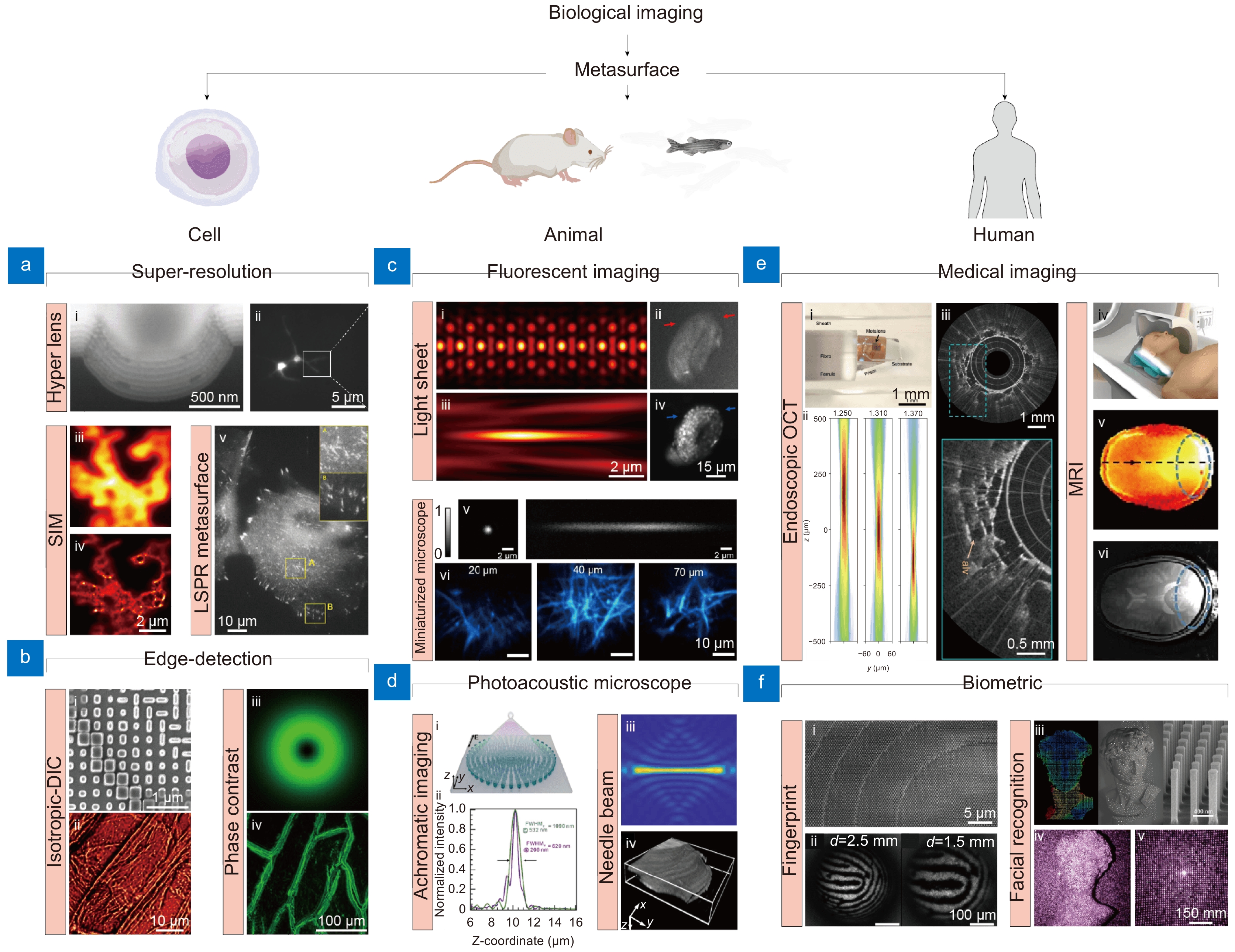
Figure 1.
Representative metasurface-based bioimaging techniques for cell, animal, and human applications. (a, b) Metasurface-based (a) super-resolution and (b) edge-detection imaging for cell imaging applications. (a-i) cross-section of hyperlens and (a-ii) neuronal super-resolution image using hyperlens. (a-iii) Diffraction-limited and (a-iv) super-resolution image of same cells using metasurface-assisted SIM, respectively. (a-v) LSPR imaging of live cell. (b-i) SEM image of edge-detection metasurface and (a-ii) i-DIC image of onion epidermal cell. (a-iii) PSF of spiral phase metasurface and (a-iv) edge-enhanced image of onion epidermal cells. (c, d) Metasurface-based (c) fluorescent and PAM for (d) animal imaging. (c i-iv) PSF of lattice light sheet microscope in (c-i) XY and (c-iii) XZ plane and in vivo fluorescent image of developing embryos obtained by (c-ii) wide field and (c-iv) light sheet microscope. (c-v) PSF of metasurface-based miniaturized two-photon microscope and (c-vi) fluorescent image of Thy1-YFP mouse brain at different depth. (d) Metasurface-based PSF engineering for PAM applications. (d-i, d-ii) Schematic and intensity profile of an achromatic metasurface for dual-channel PAM. (d-iii) needle beam PSF and (d-iv) label-free histological PAM image. (e), (f) Metasurface-based (e) medical imaging and (f) biometric devices. (e-i) Optical setup and (e-ii) PSF of metasurface-based endoscopic OCT system. (e-iii) human lung images. (e iv-vi) metasurface-assisted MRI images. (f-i) SEM image of metasurface and (f-ii) fingerprint image obtained. (f-iii) Illustration of metasurface-based point cloud generation and (f-iv, f-v) implementation of 3D facial recognition. Figure reproduced with permission from: (a) (i) ref.14, Springer Nature; (ii) ref.15 American Chemical Society; (iii, iv) ref.16, Springer Nature; (v) ref.17, American Chemical Society; (b) (i, ii) ref.18, American Chemical Society; (iii, iv) ref.19 Springer Nature; (c) (i-iii) ref.20, De Gruyter; (ii-iv) ref.21, De Gruyter; (v, vi) ref.22, American Chemical Society; (d) (i, ii) ref.23, Elsevier; (iii, iv) ref.24, Elsevier; (e) (i, iii) ref.25, Springer Nature; (iv, vi) ref.26, Springer Nature; (f) (i, ii) ref.27, American Chemical Society; (iii-v) ref.28, American Chemical Society.
-
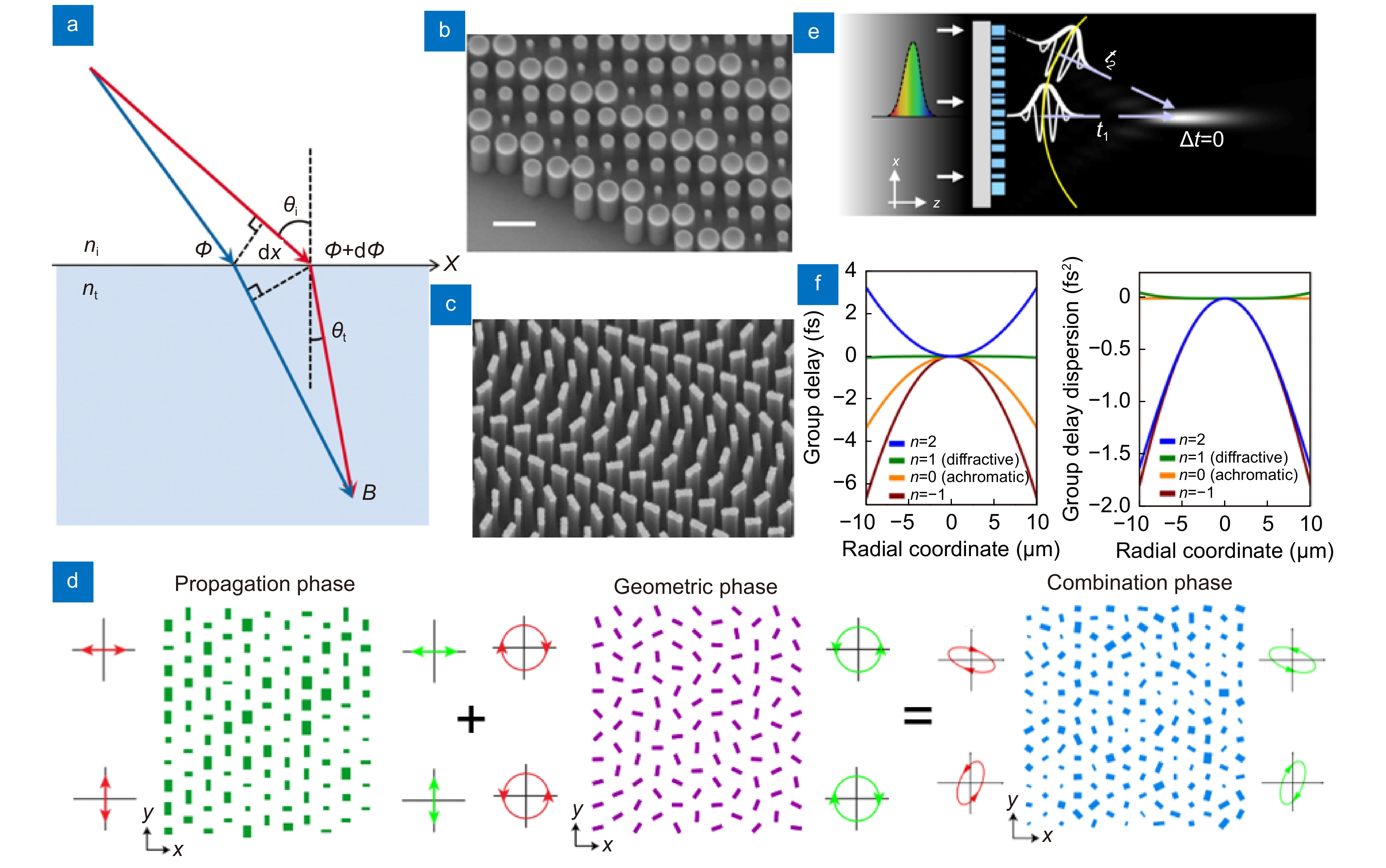
Figure 2.
Working principles of phase modulation of metasurface. (a) Schematic of the generalized Snell's law of refraction. The phase gradient causes additional refraction of light. (b) SEM image of the circular meta-atoms of propagation phase metasurface. (c) SEM image of the rectangular meta-atoms on geometric phase metasurface. (d) Schematic illustrating the principle of combining propagation and geometric phases. (e) An illustration depicting the spectral dispersion of metasurface in terms of group delay. (f) For a metasurface with a 530 nm wavelength and a 49 μm focal length, the group delay and group dispersion delay profiles are shown. Based on the value of n, the design can be optimized for achromatic or chromatic performance. Figure reproduced with permission from: (a) ref.29, American Association for the Advancement of Science; (b) ref.32, American Chemical Society; (c) ref.33, American Chemical Society; (d) adapted with permission from ref. 34, copyrighted by the American Physical Society; (e, f) ref.12, Springer Nature.
-
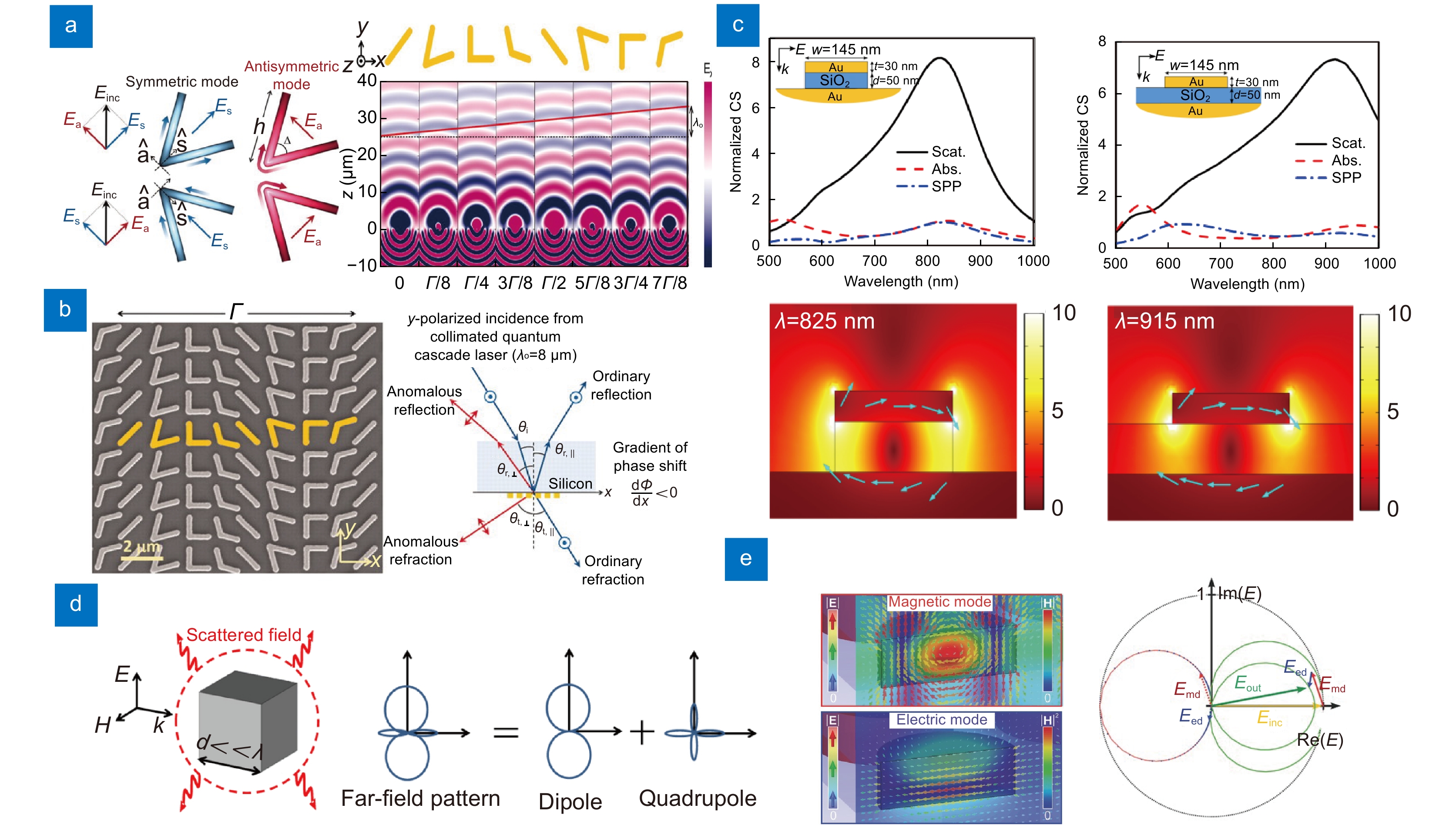
Figure 3.
Representative metasurfaces consist of plasmonic and dielectric materials. (a) V-shaped plasmonic meta-atom. The V-antennas activate symmetric and antisymmetric modes depending on the incident light. The unit cells are periodically arranged to manipulate reflection and refraction of light. (b) SEM image of V-antennas showing the periodic arrangement of unit cells (left). A schematic illustrating the phase-delayed according to the generalized Snell's law (right). (c) Scattering, absorption, and SPP cross-sections of gap surface plasmon resonator (top). Distribution of electric field enhancement (bottom). (d) A dielectric scatterer with a typical size considerably smaller than the operating wavelength. Dielectric materials are chosen for their low-loss and high refractive index properties, which allow efficient phase and amplitude control. (e) Electric and magnetic field distribution of the silicon nanodisk metasurface (left). overlapped vector diagram of the electric field (right). Electric and magnetic dipole resonances can be simultaneously generated and overlapped to suppress the reflection of the metasurface. Through the careful design of the dielectric structure, it is possible to control the transmittance using resonance control. Figure reproduced with permission from: (a, b) ref.29, American Association for the Advancement of Science; (c) ref.82, Optics express; (d) ref.88, Optics express; (e) ref.89, John Wiley and Sons.
-
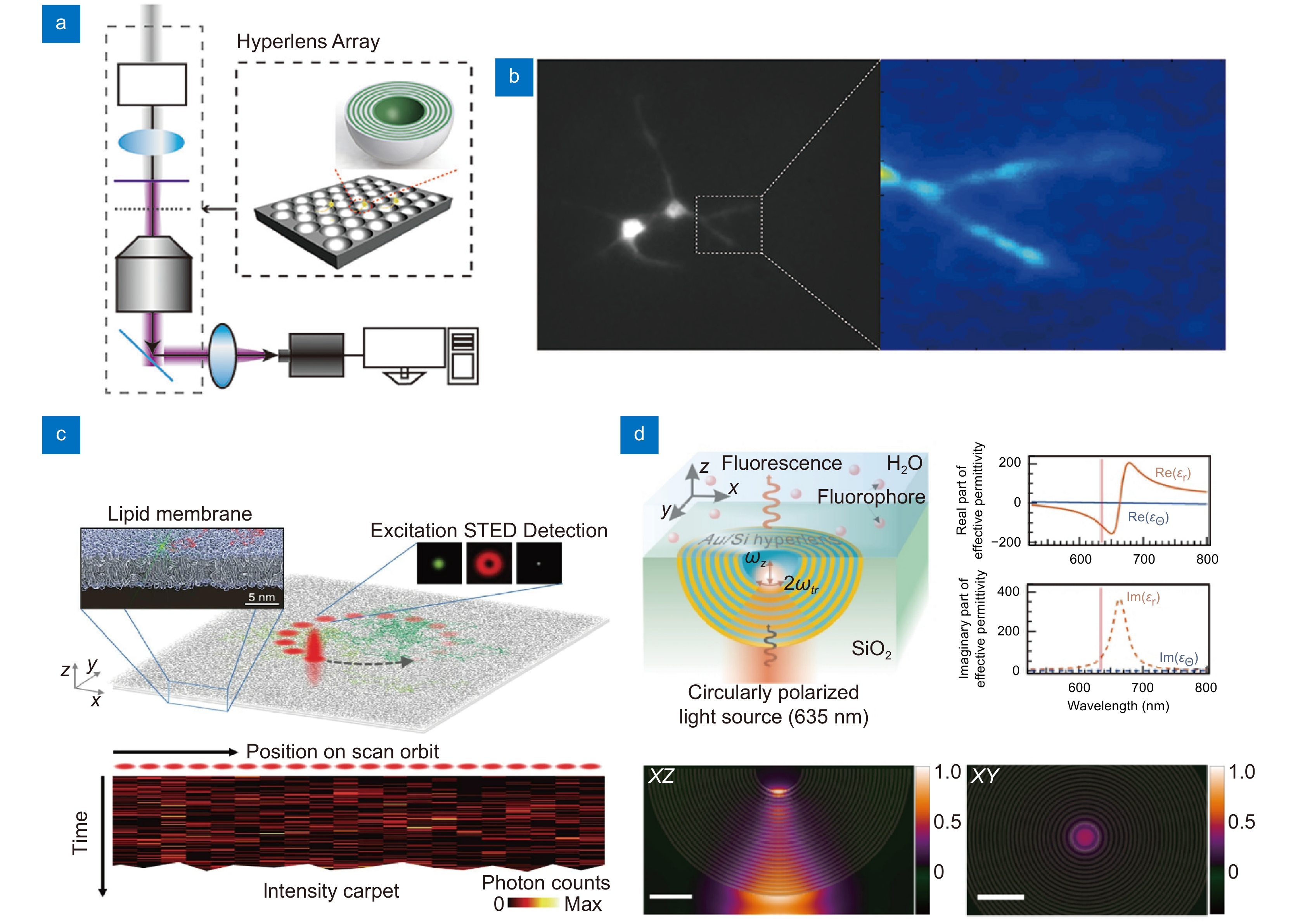
Figure 4.
Metasurfaces for super-resolution techniques for cell imaging application. (a, b) Optical setup of hyperlens-based (a) super-resolution imaging system, and (b) fluorescent image of cultured neuron. (c, d) Super-resolution FCS setup based on (c) STED, and (d) hyperlens. Scale bar: 200 nm. Figure reproduced with permission from: (a, b) ref.15, American Chemical Society; (c) ref.151, Springer Nature; (d) ref.152, Optical Society of America.
-
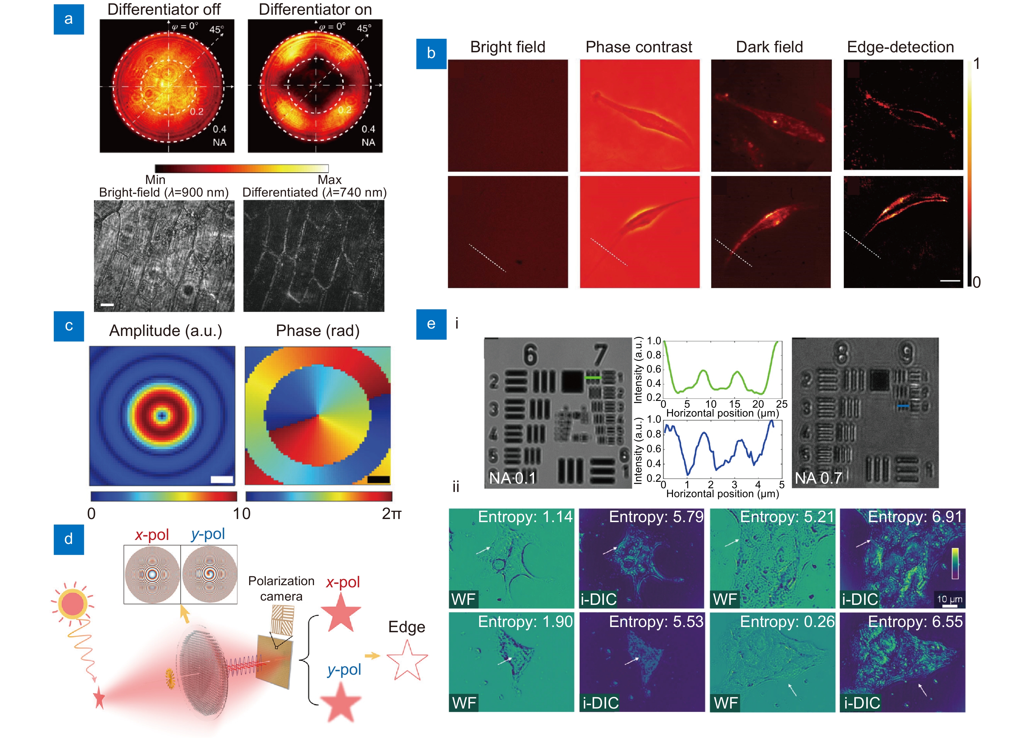
Figure 5.
Edge-detection metasurfaces for cell imaging applications. (a) Images obtained at back focal plane of a standard metasurface and nanophotonic differentiator metasurface (top). Onion epidermis obtained with and without differentiator metasurface (bottom). Scale bar: 50 μm. (b) Human umbilical vein endothelial cells (top) and human bronchial epithelial cells (middle) taken by using brightfield, phase contrast, dark field, and edge-detection, respectively. Scale bar: 100 μm. (c) Amplitude and phase distributions calculated using the angular spectrum method (ASM) at the focal plane of a spiral metasurface. Scale bar: 250 nm. (d) Edge-detection metasurface compatible to incoherent light for general purpose imaging. (e) (i) Metasurface-assisted DIC image of the USAF 1951 resolution test chart under NA of 0.1 and 0.7, respectively. (ii) Images captured with wide-field and i-DIC microscope. Figure reproduced with permission from: (a) ref.182, Springer Nature; (b) ref.186, Oxford University Press; (c) ref.188, John Wiley and Sons; (d) ref.137, American Chemical Society; (e) ref.19, Springer Nature.
-
Figure 6.
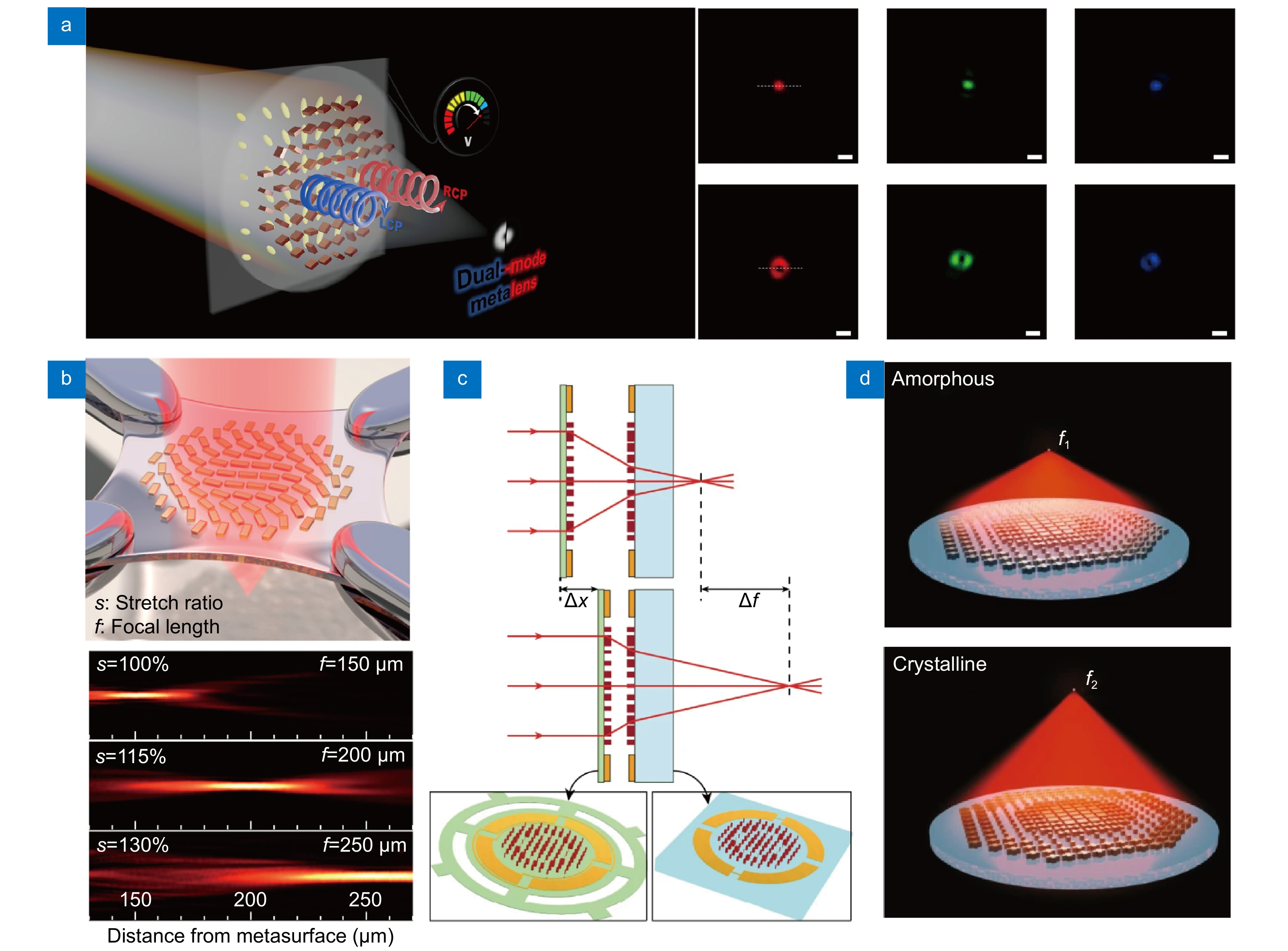
Various tunable metasurfaces and representative working principles. (a) A liquid crystal cells combined electrically tunable edge-detection metasurface. Scale bar: 5 μm. (b) Stretchable zoom metasurface fabricated on elastic substrate. (c) A MEMS combined varifocal metasurface. The focal length can be controlled by adjusting the distance be-tween two lenses using MEMS. (d) A varifocal metasurface consists of thermally responsive material that deforms in its crystalline state under heating, thereby causing a change in refractive index. Figure reproduced with permission from: (a) ref.122, American Chemical Society; (b) ref.210, American Chemical Society; (c) ref.214, Springer Nature; (d) ref.112, Springer Nature.
-
Figure 7.
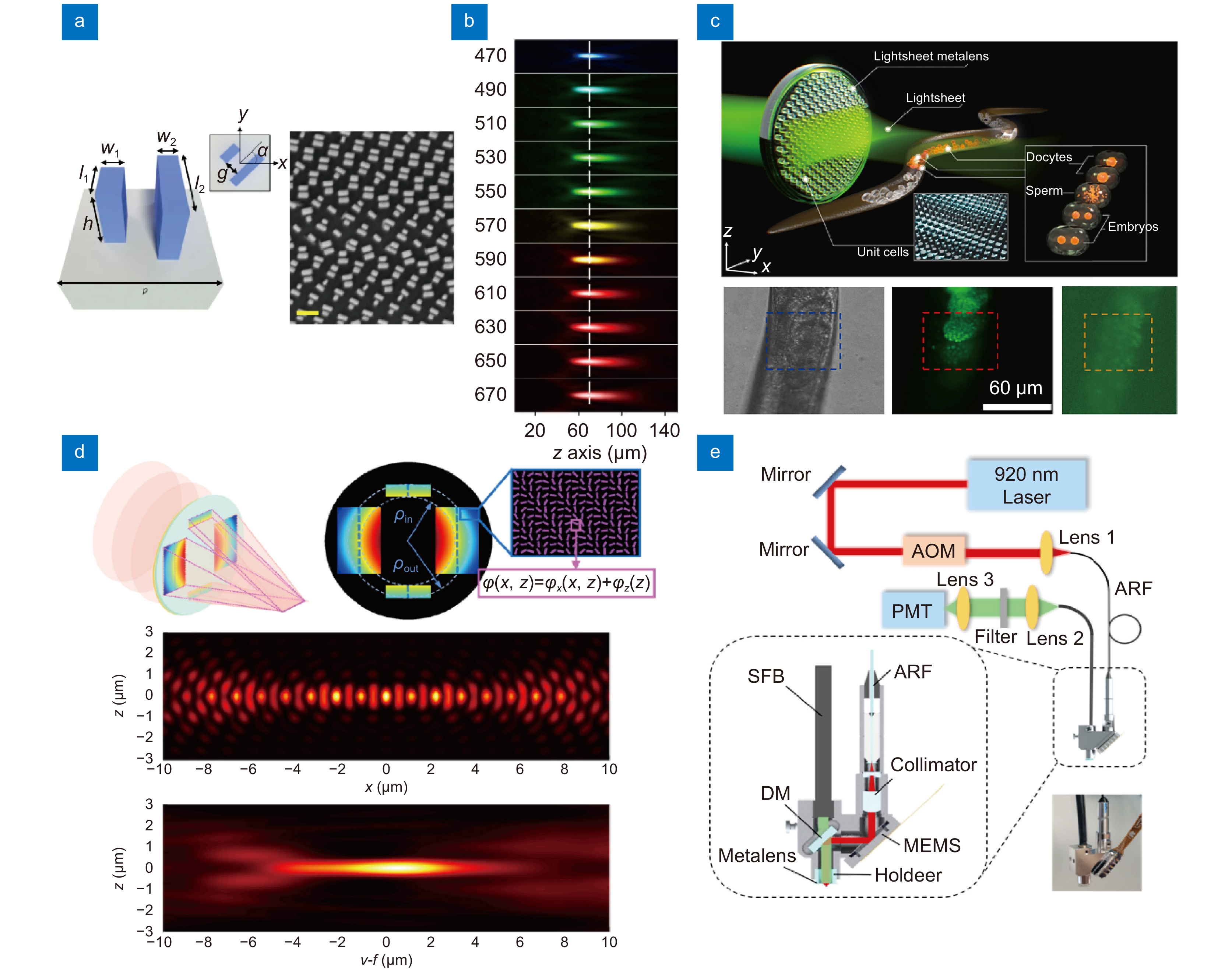
Various metasurfaces-derived fluorescent microscopies for animal imaging applications. (a) Geometry and structural parameters of unit cell consist of paired nanofins for achromatic metasurface. Scale bar: 500 nm. (b) PSF of achromatic metasurface at various wavelength. The focal plane (dotted line) coincides regardless of the wavelength. (c) Illustration of metasurface-based light sheet microscopy (top), along with a bright-field, light sheet fluorescent microscopy, and wide-field fluorescence image of C. elegans (bottom). (d) The complex phase profile of metasurface for lattice light-sheet and its PSF. (e) Miniaturized two-photon microscopy incorporating a metasurface. Figure reproduced with permission from: (a), (b) ref.12, Springer Nature; (c) ref.21, De Gruyter; (d) ref.20, De Gruyter; (e) ref.22, American Chemical Society.
-
Figure 8.
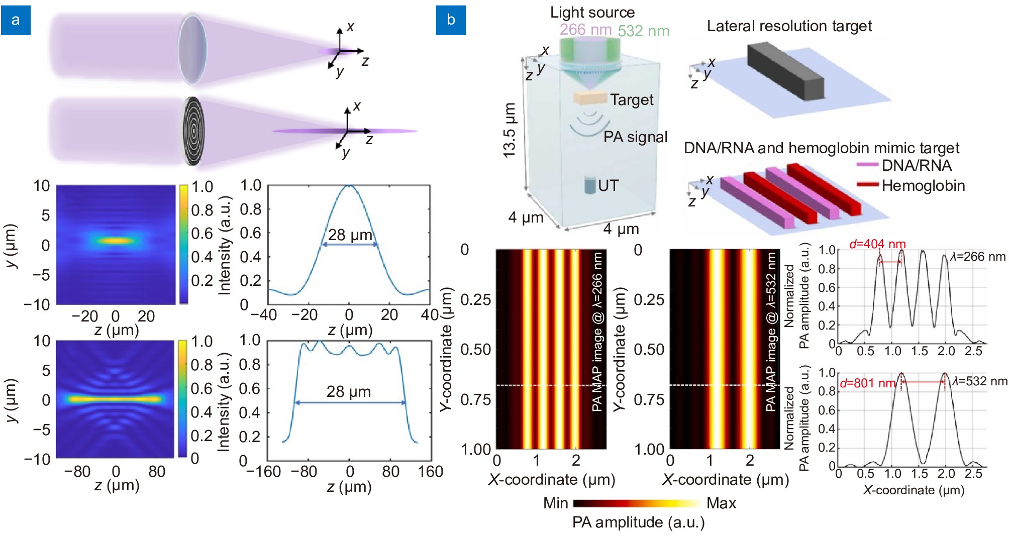
Metasurface-based PSF engineering for photoacoustic microscopy applications. (a) A comparison of PSF between a conventional objective lens with a NA of 0.13 and a metasurface designed for extended depth of focus at UV wavelength. The objective lens has a depth of focus of 28 µm, whereas the metasurface has a depth of focus of 216 µm. (b) Optical setup for dual-channel PAM using a UV-Visible metasurface (top). PAM imaging simulation and resulting intensity profiles of phantom at UV and visible light (bottom). Figure reproduced with permission from: (a) ref.244, Optical Society of America; (b) ref.23, Elsevier.
-
Figure 9.
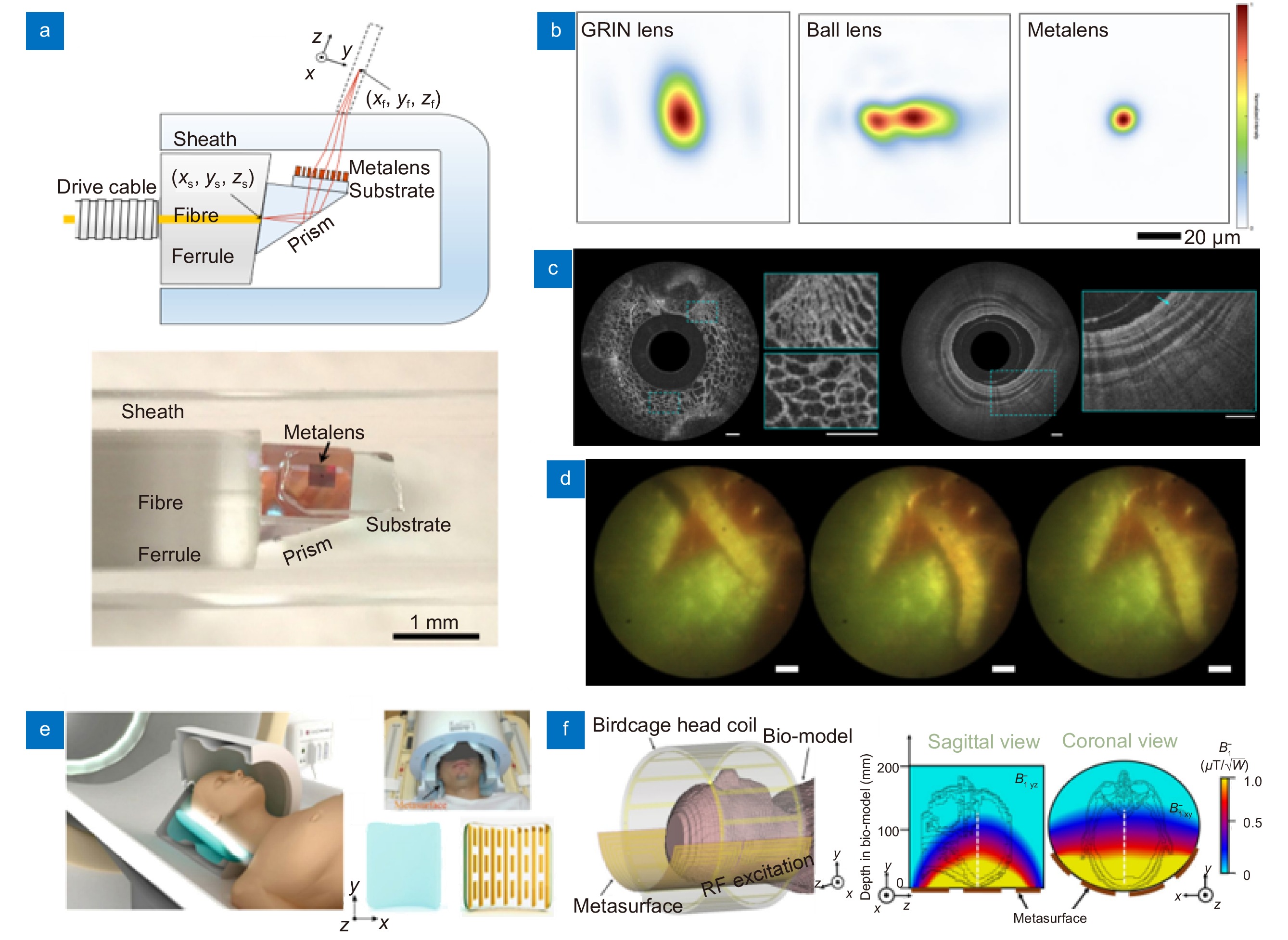
Biomedical applications of metasurfaces. (a) Schematic (top), and distal end of the nano-optic endoscope using ultra-thin metasurface. (b) Comparison of PSF at a wavelength of 1310 nm for an OCT catheter based on GRIN, ball, and metasurface, respectively. The metasurface-based OCT catheter shows the clearest PSF. (c) The image of fruit flesh of grape (left) and swine airway (right) obtained from the metasurface-based nano-optic endoscope. Scale bar: 500 μm. (d) Full color video rate imaging of caterpillar on a strawberry leaf taken from meta-optical fiber endoscope. Scale bar: 500 μm. (e) Illustration of MRI scanning with metasurface made of a high permittivity dielectric pad and metal stripes. (f) Schematic and simulation results of metasurface assisted MRI scanning. Homogeneous magnetic field enhancement is observed. Figure reproduced with permission from: (a–c) ref.25, Springer Nature; (d) ref.257, Springer Nature; (e) ref.26, Springer Nature; (f) ref.263, Springer Nature.
-
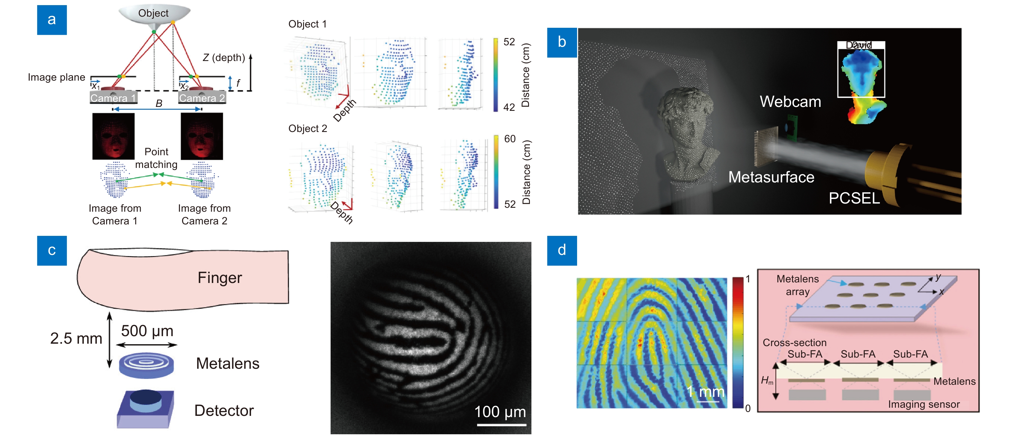
Figure 10.
Representative metasurface integrated devices for biometric applications. (a) Implementation of 3D facial depth sensing using a point cloud generating metasurface. (b) Illustration of a metasurface-based dot projector combined with PCSEL, which is 233 times smaller than a commercial DOE-based dot projector. (c) Schematic of metasurface-based device for fingerprint recognition (left) and fingerprint image obtained by the device. (d) Fingerprint image obtained using a metasurface array imaging device. The array structure enables the acquisi-tion of clearer and wider fingerprint images. Figure reproduced with permission from: (a) ref.49, Springer Nature; (b) ref.28, American Chemical Society; (c) ref.27, American Chemical Society; (d) ref.283, De Gruyter.

 E-mail Alert
 RSS
RSS

 DownLoad:
