
 E-mail Alert
 RSS
RSS
| Citation: |
Wei XR, Liang YZ, Zhang XH et al. Multi-resonance enhanced photothermal synergistic fiber-optic Tamm plasmon polariton tip for high-sensitivity and rapid hydrogen detection. Opto-Electron Sci 4, 240029 (2025). doi: 10.29026/oes.2025.240029

|
Multi-resonance enhanced photothermal synergistic fiber-optic Tamm plasmon polariton tip for high-sensitivity and rapid hydrogen detection
-
Abstract
Accurate and real-time detection of hydrogen (H2) is essential for ensuring energy security. Fiber-optic H2 sensors are gaining attention for their integration and remote sensing capabilities. However, they face challenges, including complex fabrication processes and limited response times. Here, we propose a fiber-optic H2 sensing tip based on Tamm plasmon polariton (TPP) resonance, consisting of a multilayer metal/dielectric Bragg reflector deposited directly on the fiber end facet, simplifying the fabrication process. The fiber-optic TPP (FOTPP) tip exhibits both TPP and multiple Fabry-Perot (FP) resonances simultaneously, with the TPP employed for highly sensitive H2 detection. Compared to FP resonance, TPP exhibits more than twice the sensitivity under the same structural dimension without cavity geometry deformation. The excellent performance is attributed to alterations in phase-matching conditions, driven by changes in penetration depth of TPP. Furthermore, the FP mode is utilized to achieve an efficient photothermal effect to catalyze the reaction between H2 and the FOTPP structure. Consequently, the response and recovery speeds of the FOTPP tip under resonance-enhanced photothermal assistance are improved by 6.5 and 2.1 times, respectively. Our work offers a novel strategy for developing TPP-integrated fiber-optic tips, refines the theoretical framework of photothermal-assisted detection systems, and provides clear experimental evidence. -

-
References
[1] Ould Amrouche S, Rekioua D, Rekioua T et al. Overview of energy storage in renewable energy systems. Int J Hydrogen Energy 41, 20914–20927 (2016). doi: 10.1016/j.ijhydene.2016.06.243 [2] Yue ML, Lambert H, Pahon E et al. Hydrogen energy systems: a critical review of technologies, applications, trends and challenges. Renew Sustain Energy Rev 146, 111180 (2021). doi: 10.1016/j.rser.2021.111180 [3] Sripriya, Meda US. Market study of hydrogen sensors and sensing systems. ECS Trans 107, 4489–4502 (2022). doi: 10.1149/10701.4489ecst [4] Tittl A, Mai P, Taubert R et al. Palladium-based plasmonic perfect absorber in the visible wavelength range and its application to hydrogen sensing. Nano Lett 11, 4366–4369 (2011). doi: 10.1021/nl202489g [5] Baldi A, Narayan TC, Koh AL et al. In situ detection of hydrogen-induced phase transitions in individual palladium nanocrystals. Nat Mater 13, 1143–1148 (2014). doi: 10.1038/nmat4086 [6] Matuschek M, Singh DP, Jeong HH et al. Chiral plasmonic hydrogen sensors. Small 14, 1702990 (2018). doi: 10.1002/smll.201702990 [7] Wen L, Sun ZW, Zheng QL et al. On-chip ultrasensitive and rapid hydrogen sensing based on plasmon-induced hot electron–molecule interaction. Light Sci Appl 12, 76 (2023). doi: 10.1038/s41377-023-01123-4 [8] Tomeček D, Moberg HK, Nilsson S et al. Neural network enabled nanoplasmonic hydrogen sensors with 100 ppm limit of detection in humid air. Nat Commun 15, 1208 (2024). doi: 10.1038/s41467-024-45484-9 [9] Korotcenkov G, Han SD, Stetter JR. Review of electrochemical hydrogen sensors. Chem Rev 109, 1402–1433 (2009). doi: 10.1021/cr800339k [10] Liu Q, Yao JY, Wang YP et al. Temperature dependent response/recovery characteristics of Pd/Ni thin film based hydrogen sensor. Sens Actuators B Chem 290, 544–550 (2019). doi: 10.1016/j.snb.2019.04.024 [11] Caucheteur C, Guo T, Albert J. Review of plasmonic fiber optic biochemical sensors: improving the limit of detection. Anal Bioanal Chem 407, 3883–3897 (2015). doi: 10.1007/s00216-014-8411-6 [12] Jing JY, Liu K, Jiang JF et al. Highly sensitive and stable probe refractometer based on configurable plasmonic resonance with nano-modified fiber core. Opto-Electron Adv 6, 220072 (2023). doi: 10.29026/oea.2023.220072 [13] Wang Q, Wang L. Lab-on-fiber: plasmonic nano-arrays for sensing. Nanoscale 12, 7485–7499 (2020). doi: 10.1039/D0NR00040J [14] Xiong YF, Xu F. Multifunctional integration on optical fiber tips: challenges and opportunities. Adv Photonics 2, 064001 (2020). [15] Zhao Y, Tong RJ, Xia F et al. Current status of optical fiber biosensor based on surface plasmon resonance. Biosens Bioelectron 142, 111505 (2019). doi: 10.1016/j.bios.2019.111505 [16] Liu HH, Hu DJJ, Sun QZ, Wei L, Li KW et al. Specialty optical fibers for advanced sensing applications. Opto-Electron Sci 2, 220025 (2023). doi: 10.29026/oes.2023.220025 [17] Jiang BQ, Hou YG, Wu JX, Ma YX, Gan XT et al. In-fiber photoelectric device based on graphene-coated tilted fiber grating. Opto-Electron Sci 2, 230012 (2023). doi: 10.29026/oes.2023.230012 [18] Yin SY, Guo Q, Liu SR et al. Three-dimensional multichannel waveguide grating filters. Opto-Electron Sci 3, 240003 (2024). doi: 10.29026/oes.2024.240003 [19] Zhang YN, Liu YX, Shi BF et al. Lateral offset single-mode fiber-based Fabry–Perot interferometers with Vernier effect for hydrogen sensing. Anal Chem 95, 872–880 (2023). [20] Luo JX, Liu S, Chen PJ et al. Highly sensitive hydrogen sensor based on an optical driven nanofilm resonator. ACS Appl Mater Interfaces 14, 29357–29365 (2022). doi: 10.1021/acsami.2c04105 [21] Wang CQ, Han ZW, Wang CX et al. Highly sensitive fiber grating hydrogen sensor based on hydrogen-doped Pt/WO3. Sens Actuators B Chem 404, 135250 (2024). doi: 10.1016/j.snb.2023.135250 [22] Ye Z, Ruan HB, Hu XY et al. TBAOH intercalated WO3 for high-performance optical fiber hydrogen sensor. Int J Hydrogen Energy 47, 28204–28211 (2022). doi: 10.1016/j.ijhydene.2022.06.133 [23] Cai SS, Liu F, Wang RL et al. Narrow bandwidth fiber-optic spectral combs for renewable hydrogen detection. Sci China Inf Sci 63, 222401 (2020). doi: 10.1007/s11432-020-3058-2 [24] Gu FX, Wu GQ, Zeng HP. Hybrid photon–plasmon Mach–Zehnder interferometers for highly sensitive hydrogen sensing. Nanoscale 7, 924–929 (2015). doi: 10.1039/C4NR06642A [25] Perrotton C, Westerwaal RJ, Javahiraly N et al. A reliable, sensitive and fast optical fiber hydrogen sensor based on surface plasmon resonance. Opt Express 21, 382–390 (2013). doi: 10.1364/OE.21.000382 [26] Nugroho FAA, Eklund R, Nilsson S et al. A fiber-optic nanoplasmonic hydrogen sensor via pattern-transfer of nanofabricated PdAu alloy nanostructures. Nanoscale 10, 20533–20539 (2018). doi: 10.1039/C8NR03751E [27] Yun S, Oyama ST. Correlations in palladium membranes for hydrogen separation: a review. J Memb Sci 375, 28–45 (2011). doi: 10.1016/j.memsci.2011.03.057 [28] Adhikari S, Efremova MV, Spaeth P et al. Single-particle photothermal circular dichroism and photothermal magnetic circular dichroism microscopy. Nano Lett 24, 5093–5103 (2024). doi: 10.1021/acs.nanolett.4c00448 [29] Cui XM, Ruan QF, Zhuo XL et al. Photothermal nanomaterials: a powerful light-to-heat converter. Chem Rev 123, 6891–6952 (2023). doi: 10.1021/acs.chemrev.3c00159 [30] Luo J, Wu QL, Zhou L et al. Plasmon-induced hot carrier dynamics and utilization. Photonics Insights 2, R08 (2023). doi: 10.3788/PI.2023.R08 [31] Polley N, Sardar S, Werner P et al. Photothermomechanical nanopump: a flow-through plasmonic sensor at the fiber tip. ACS Nano 17, 1403–1413 (2023). doi: 10.1021/acsnano.2c09938 [32] Stewart JW, Nebabu T, Mikkelsen MH. Control of nanoscale heat generation with lithography-free metasurface absorbers. Nano Lett 22, 5151–5157 (2022). doi: 10.1021/acs.nanolett.2c00761 [33] Wu HT, Chen PW, Zhan XD et al. Marriage of a dual-plasmonic interface and optical microfiber for NIR-II cancer theranostics. Adv Mater 36, 2310571 (2024). doi: 10.1002/adma.202310571 [34] Ye Z, Li Z, Dai JX et al. Hydrogen sensing performance investigations with optical heating and sensing element surface modification. Int J Hydrogen Energy 46, 1411–1419 (2021). doi: 10.1016/j.ijhydene.2020.09.140 [35] Zhang XP, Li XT, Zhang XH et al. Photothermal-assisted hydrogen permeation enhancement. Sens Actuators B Chem 365, 131935 (2022). doi: 10.1016/j.snb.2022.131935 [36] Kaliteevski M, Iorsh I, Brand S et al. Tamm plasmon-polaritons: possible electromagnetic states at the interface of a metal and a dielectric Bragg mirror. Phys Rev B 76, 165415 (2007). doi: 10.1103/PhysRevB.76.165415 [37] Lundt N, Klembt S, Cherotchenko E et al. Room-temperature Tamm-plasmon exciton-polaritons with a WSe2 monolayer. Nat Commun 7, 13328 (2016). doi: 10.1038/ncomms13328 [38] Hu MY, Zhang Y, Jiang X et al. Double-bowl state in photonic Dirac nodal line semimetal. Light Sci Appl 10, 170 (2021). doi: 10.1038/s41377-021-00614-6 [39] Kar C, Jena S, Udupa DV et al. Tamm plasmon polariton in planar structures: a brief overview and applications. Opt Laser Technol 159, 108928 (2023). doi: 10.1016/j.optlastec.2022.108928 [40] Normani S, Bertolotti P, Bisio F et al. Tamm plasmon resonance as optical fingerprint of silver/bacteria interaction. ACS Appl Mater Interfaces 15, 27750–27758 (2023). doi: 10.1021/acsami.3c05473 [41] Sreekanth KV, Perumal J, Dinish US et al. Tunable Tamm plasmon cavity as a scalable biosensing platform for surface enhanced resonance Raman spectroscopy. Nat Commun 14, 7085 (2023). doi: 10.1038/s41467-023-42854-7 [42] He MZ, Nolen JR, Nordlander J et al. Coupled Tamm phonon and plasmon polaritons for designer planar multiresonance absorbers. Adv Mater 35, 2209909 (2023). doi: 10.1002/adma.202209909 [43] Wang ZY, Ho YL, Cao T et al. High-Q and tailorable fano resonances in a one-dimensional metal-optical Tamm state structure: from a narrowband perfect absorber to a narrowband perfect reflector. Adv Funct Mater 31, 2102183 (2021). doi: 10.1002/adfm.202102183 [44] Lu H, Shi SH, Li DK et al. Strong self-enhancement of optical nonlinearity in a topological insulator with generation of Tamm state. Laser Photonics Rev 17, 2300269 (2023). doi: 10.1002/lpor.202300269 [45] Ko JH, Seo DH, Jeong HH et al. Sub-1-volt electrically programmable optical modulator based on active Tamm plasmon. Adv Mater 36, 2310556 (2024). doi: 10.1002/adma.202310556 [46] Zhang KH, Chen ZY, Li HJ et al. A dual-band hydrogen sensor based on Tamm plasmon polaritons. Phys Chem Chem Phys 25, 20697–20705 (2023). doi: 10.1039/D3CP02653A [47] Palm KJ, Murray JB, Narayan TC et al. Dynamic optical properties of metal hydrides. ACS Photonics 5, 4677–4686 (2018). doi: 10.1021/acsphotonics.8b01243 [48] Brovelli LR, Keller U. Simple analytical expressions for the reflectivity and the penetration depth of a Bragg mirror between arbitrary media. Opt Commun 116, 343–350 (1995). doi: 10.1016/0030-4018(95)00084-L [49] Adams M, Cemlyn B, Henning I et al. Model for confined Tamm plasmon devices. J Opt Soc Am B 36, 125–130 (2019). doi: 10.1364/JOSAB.36.000125 [50] Peleg M, Normand MD, Corradini MG. The Arrhenius equation revisited. Crit Rev Food Sci Nutr 52, 830–851 (2012). doi: 10.1080/10408398.2012.667460 [51] Chen X, Chen YT, Yan M et al. Nanosecond photothermal effects in plasmonic nanostructures. ACS Nano 6, 2550–2557 (2012). doi: 10.1021/nn2050032 -
Supplementary Information
Supplementary information for Multi-resonance enhanced photothermal synergistic fiber-optic Tamm plasmon polariton tip for high-sensitivity and rapid hydrogen detection 
-
Access History
Figures(6)
Tables(2)
Article Metrics


Export File
Citation
Wei XR, Liang YZ, Zhang XH et al. Multi-resonance enhanced photothermal synergistic fiber-optic Tamm plasmon polariton tip for high-sensitivity and rapid hydrogen detection. Opto-Electron Sci 4, 240029 (2025). doi: 10.29026/oes.2025.240029
Format
Content
 DownLoad:
-
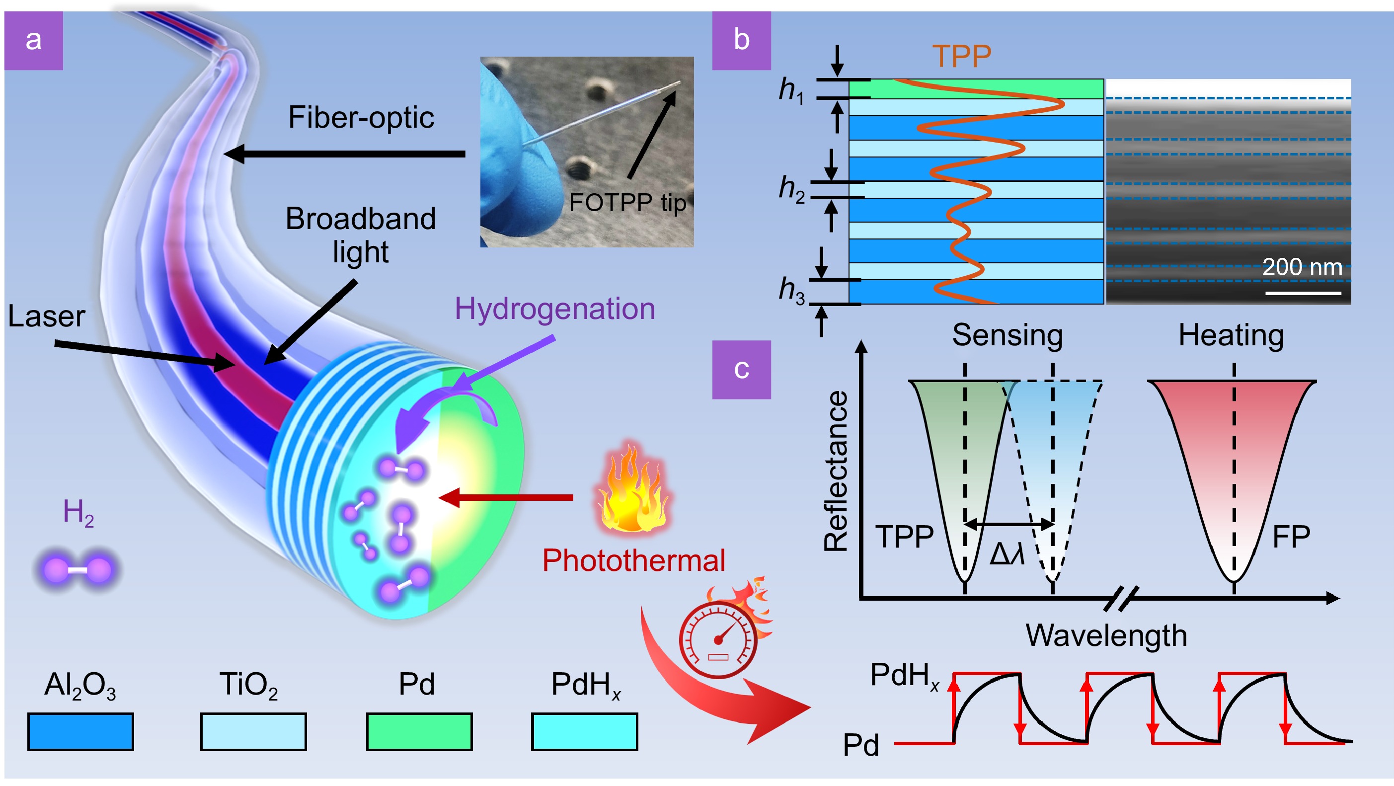
Figure 1.
Photothermal synergistic TPP H2 detection system integrated on the fiber tip. (a) Three-dimensional schematic of the fabricated fiber-optic TPP (FOTPP) tip with broadband halogen source (blue beam) and narrowband laser (red beam), along with a photograph of the actual fabricated FOTPP tip. (b) Its cross-section and SEM image for the FOTPP tip, where h1, h2, and h3 denote the thicknesses of Pd, TiO2, and Al2O3 layers, respectively. (c) Schematic of spectral working principle of photothermal synergistic FOTPP H2 sensing tip and the response/recovery characteristics of resonance wavelength under photothermal (red curve) and non-photothermal (black curve) synergistic conditions.
-
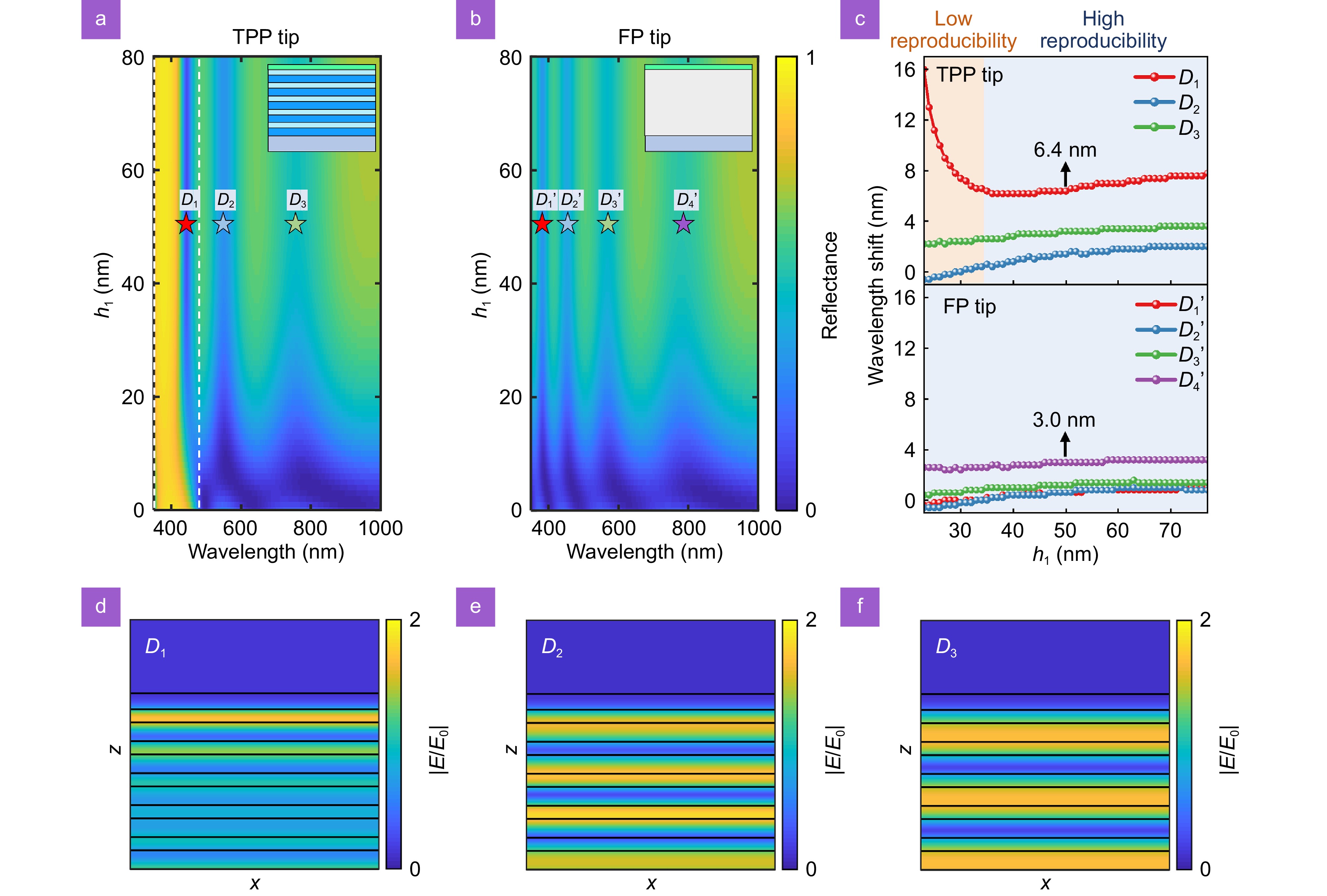
Figure 2.
Photoelectric characteristics of the FOTPP tip for H2 detection. TMM calculated the reflection spectra of (a) FOTPP and (b) FP tips with different thicknesses of Pd film. The white dashed line marks the main bandgap location of the DBR. (c) Wavelength shifts of the resonance dips of TPP (top panel) and FP (bottom panel) tips after hydrogenation at various thicknesses of Pd film. (d–f) Simulated electric field profiles for three resonance dips supported by the FOTPP tip with a 50 nm thick Pd film, where E0 denotes the incident electric field intensity.
-
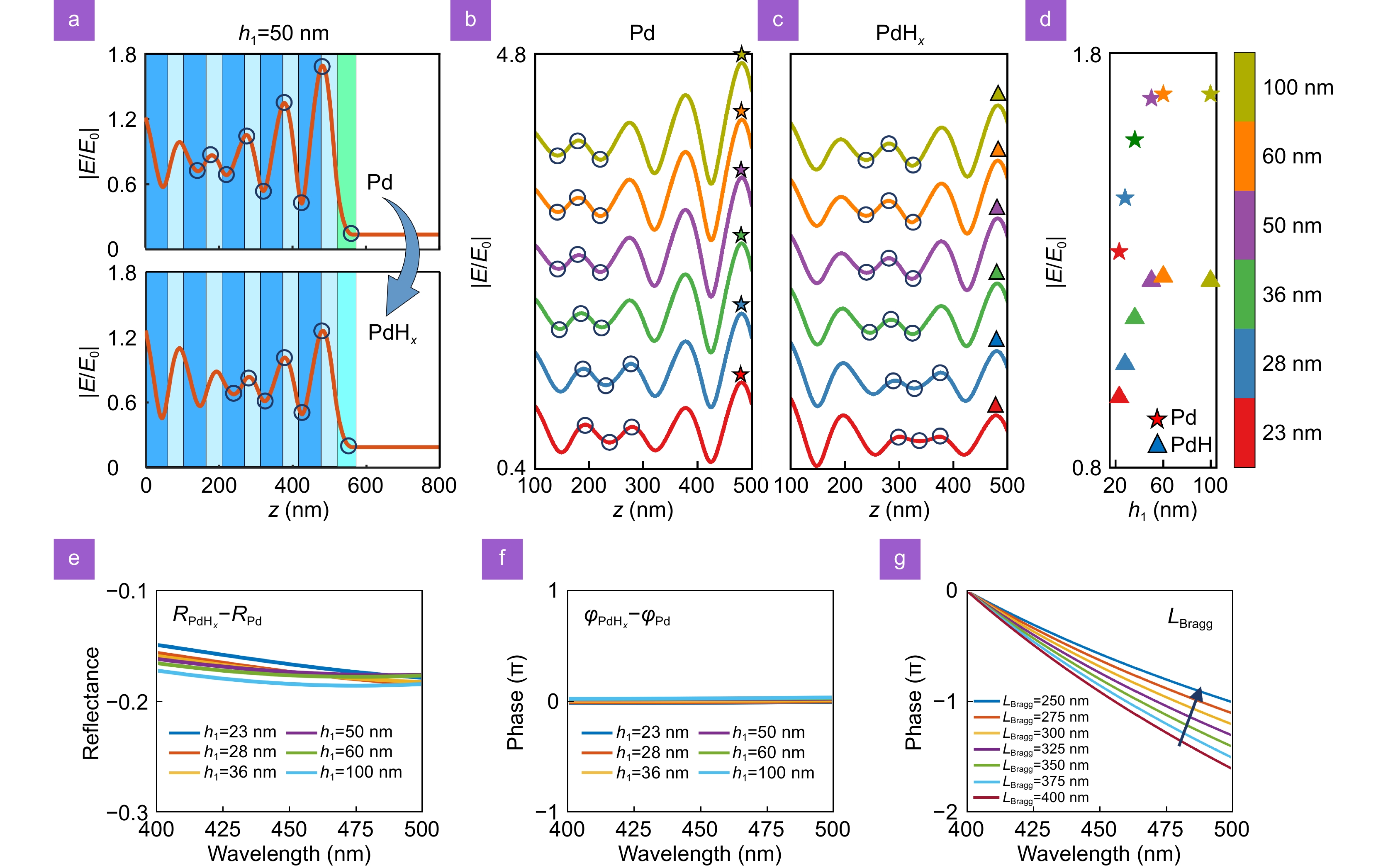
Figure 3.
Response mechanisms of resonance wavelength in TPP mode. (a) Calculated electric field distribution at the resonance wavelength of TPP supported by the FOTPP tip before (top panel) and after hydrogenation (bottom panel). The dependence of the electric field distribution of TPP (b) before and (c) after hydrogenation on the thickness of the Pd film. The individual electric field curves are shifted upwards for clarity. The circle marks indicate the position range of LBragg (near the minimum of the electric field envelope). (d) The maximum electric field intensity of TPP at the Pd/DBR interface as a function of Pd film thickness. (e) Reflection intensity difference and (f) reflection phase difference between PdHx and Pd films at different thicknesses. (g) Dependency of the total phase contribution of the DBR on the penetration depth of TPP in DBR (LBragg).
-
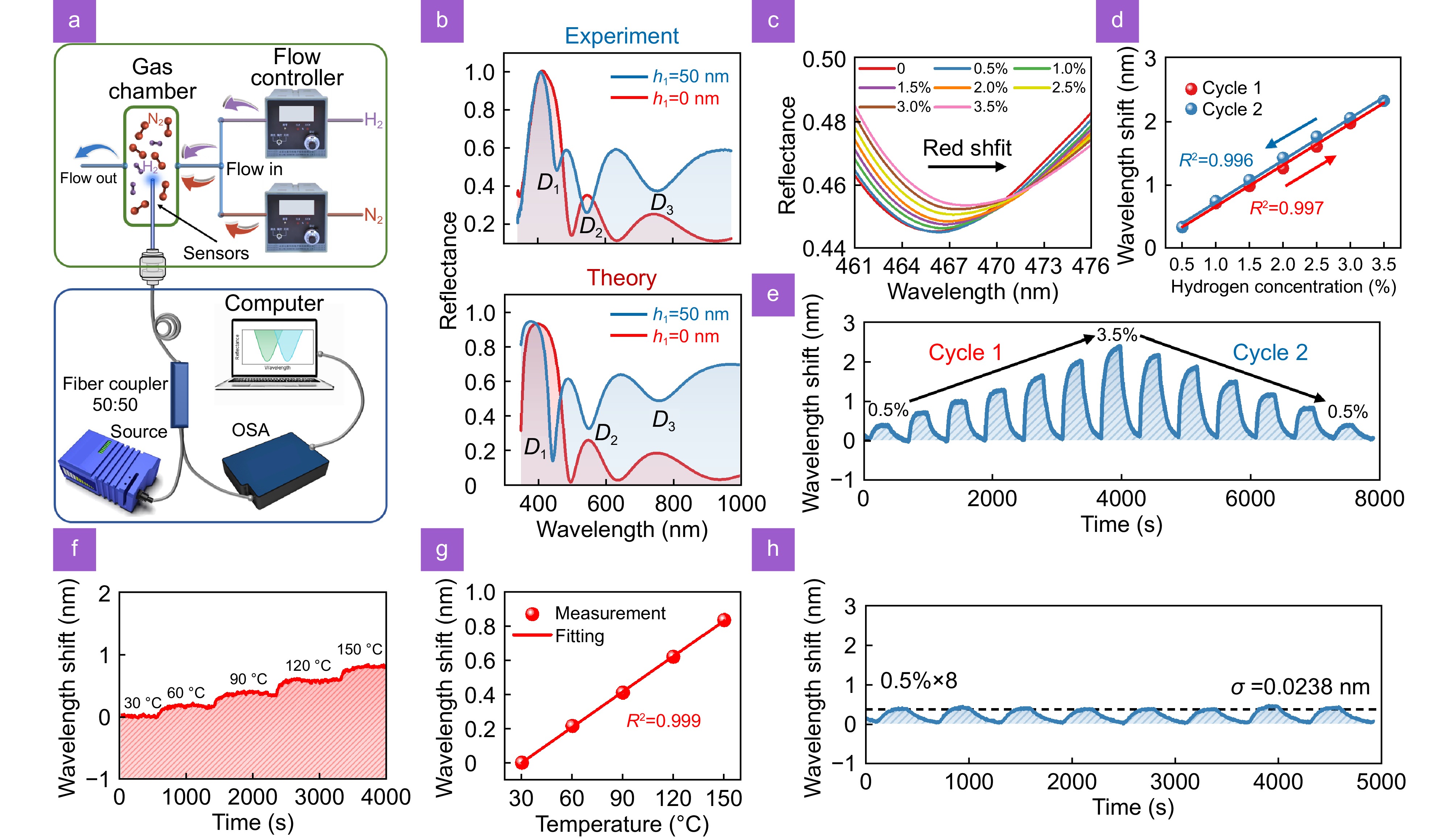
Figure 4.
Experimental measurements of H2 concentration using the fabricated FOTPP tip. (a) Schematic of the experimental setup for H2 detection, highlighting the gas sensing and spectral analysis systems in the green and blue boxes, respectively. (b) Experimentally measured (top panel) and theoretically calculated (bottom panel) reflection spectra of the FOTPP tip without a Pd film and with a 50 nm thick Pd film. (c) Reflection spectra of the FOTPP tip under various H2 concentrations ranging from 0.5% to 3.5%. (d) Wavelength redshifts and (e) real-time response of wavelength shift for the FOTPP tip at both increasing and decreasing H2 concentration pulses, ranging from 0.5% to 3.5% and 3.5% to 0.5%. (f) Real-time wavelength shift response of TPP supported by the FOTPP tip under different working temperatures. (g) The relationship between the amount of wavelength shifts of TPP and working temperatures. (h) The wavelength response of the FOTPP tip in continuously repeated 0.5% H2 concentration. The black dashed line represents the locations of the average wavelength shifts.
-
Figure 5.
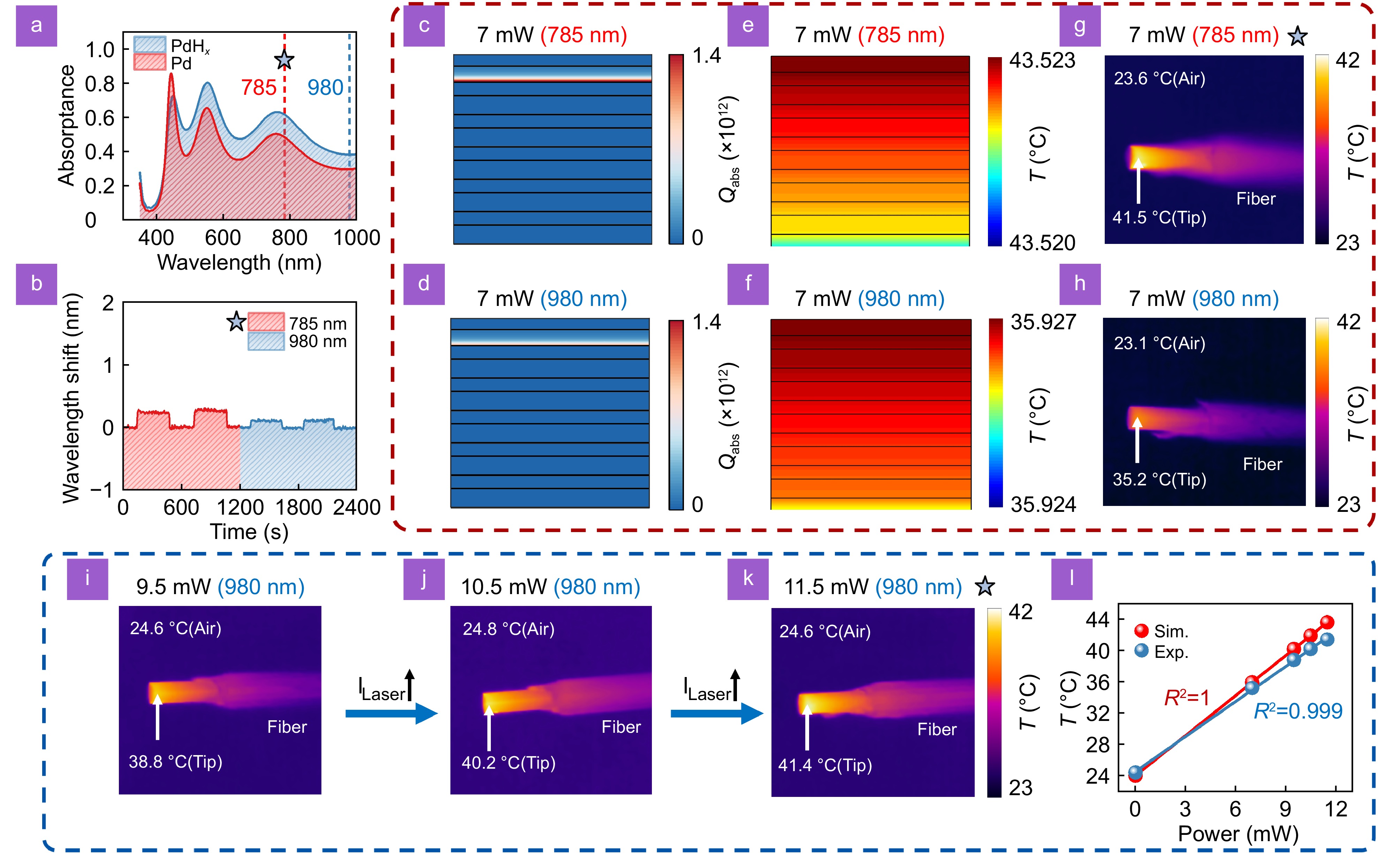
Theoretical and experimental characterization of photothermal effect in the FOTPP tip with a 50 nm thick Pd film. (a) Simulated absorption spectra of the FOTPP tip before and after hydrogenation. (b) The effect of two 7 mW lasers with different wavelengths on the resonance wavelength of TPP. Calculated absorbed power and temperature distributions at wavelengths of (c, d) 785 nm and (e, f) 980 nm. Experimentally measured thermal images of the FOTPP tip under (g) 785 nm and (h) 980 nm laser illumination. (i–k) Thermal images of the FOTPP tip illuminated with three higher-power 985 nm lasers (9.5 mW, 10.5 mW, and 11.5 mW). The peak temperature of the fiber-optic tip and the ambient temperature are indicated in the thermal images. (l) Simulated and measured peak temperature at the FOTPP tip under different laser powers. For clarity, scenarios illuminated by 7 mW and higher power lasers are differentiated by enclosing them in red and blue boxes, respectively.
-
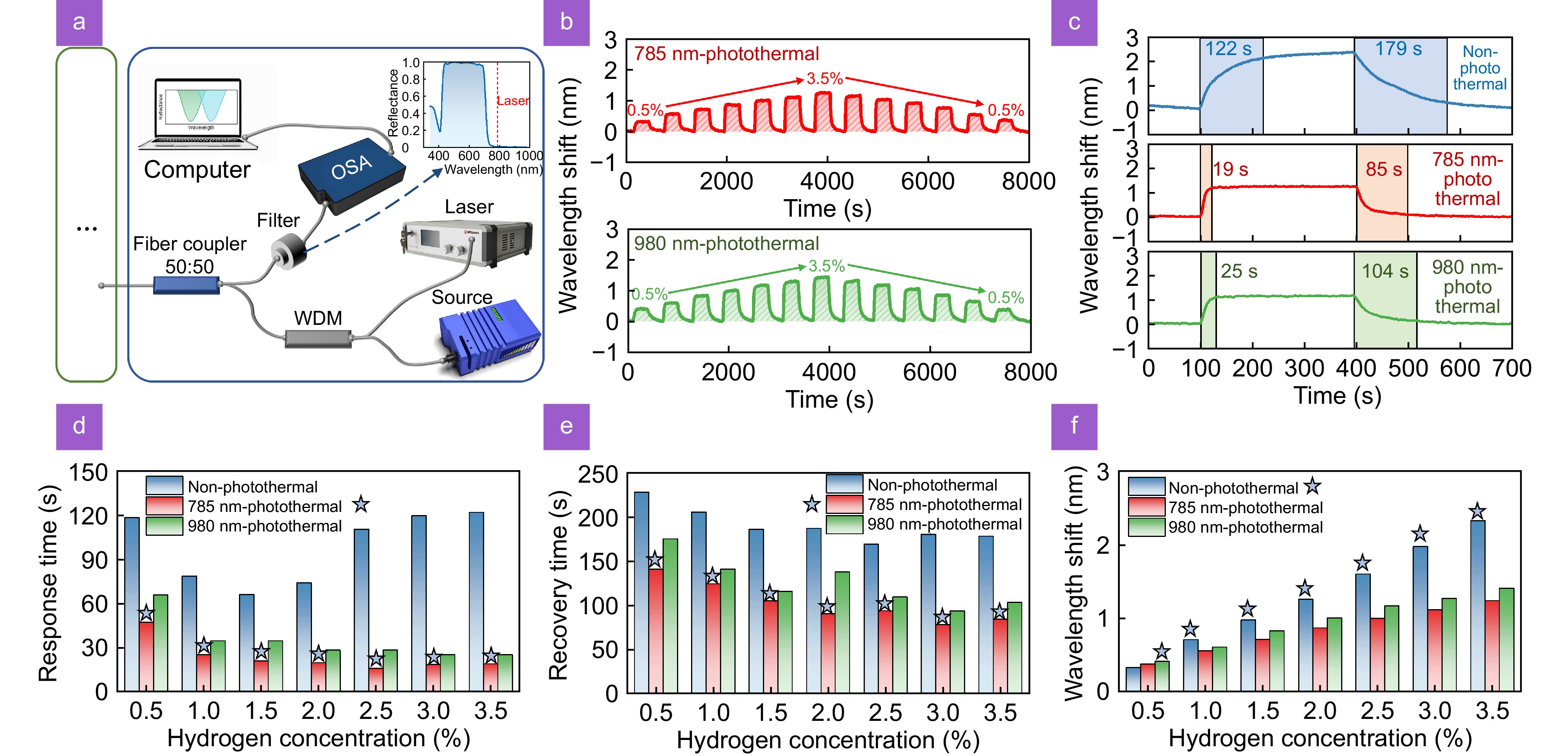
Figure 6.
Quantitatively assessment of the sensing performance of resonance-enhanced photothermal synergistic FOTPP tip with a 50 nm thick Pd film. (a) Schematic of the optical setup used for photothermal assistance. (b) Real-time wavelengths shift of TPP mode supported by the FOTPP tip under 785 nm laser illumination (top panel) and 980 nm laser illumination (bottom panel). (c) Response recovery curve of TPP at a 3.5% H2 concentration under non-photothermal (blue curve), 785 nm-photothermal (red curve), and 980 nm-photothermal (green curve) conditions. Comparison of (d) response time, (e) recovery time, and (f) wavelength redshifts for the FOTPP tip at various H2 concentrations under three different photothermal conditions.


