
 E-mail Alert
 RSS
RSS
| Citation: |
Yin Z D, Ni C D, Wu S Z, et al. Femtosecond laser direct writing processing of SERS substrates and applications[J]. Opto-Electron Eng, 2023, 50(3): 220322. doi: 10.12086/oee.2023.220322

|
Femtosecond laser direct writing processing of SERS substrates and applications
-
Abstract
Surface-enhanced Raman spectroscopy (SERS) technique plays an important role in molecular recognition fields due to its highly sensitive and high-resolution. As an emerging low-cost, high machining accuracy, and high-flexibility processing method, femtosecond laser direct writing processing has been widely used in the field of preparing SERS substrates. This work introduces four methods of preparing SERS substrates by femtosecond laser direct writing, including femtosecond two-photon reduction, femtosecond laser cutting metal, femtosecond laser cutting-sputtering, and femtosecond laser 3D printing. The article introduces the performance and application scenarios of each method in preparing SERS substrates and illustrates the advantages of femtosecond laser direct writing processing in preparing SERS substrates, aiming to provide a reference for future related research.-
Keywords:
- SERS /
- femtosecond laser direct writing /
- micro/nano processing /
- SERS substrate
-

-
References
[1] Zong C, Xu M X, Xu L J, et al. Surface-enhanced Raman spectroscopy for Bioanalysis: reliability and challenges[J]. Chem Rev, 2018, 118(10): 4946−4980. doi: 10.1021/acs.chemrev.7b00668 [2] Xu K C, Wang Z Y, Tan C F, et al. Uniaxially stretched flexible surface Plasmon resonance film for versatile surface enhanced Raman scattering diagnostics[J]. ACS Appl Mater Interfaces, 2017, 9(31): 26341−26349. doi: 10.1021/acsami.7b06669 [3] Xu K C, Zhou R, Takei K, Hong M H. Toward flexible surface-enhanced Raman scattering (SERS) sensors for point-of-care diagnostics[J]. Adv Sci (Weinh), 2019, 6(16): 1900925. doi: 10.1002/advs.201900925 [4] Niu R J, Gao F, Wang D, et al. Pattern recognition directed assembly of Plasmonic gap nanostructures for single-molecule SERS[J]. ACS Nano, 2022, 16(9): 14622−14631. doi: 10.1021/acsnano.2c05150 [5] Lee S, Jung I, Son J, et al. Heterogeneous component Au (Outer)-Pt (Middle)-Au (Inner) Nanorings: synthesis and vibrational characterization on middle Pt Nanorings with surface-enhanced Raman scattering[J]. ACS Nano, 2022, 16(7): 11259−11267. doi: 10.1021/acsnano.2c04633 [6] Qin M, Ge M H, Li P, et al. Natural <3 nm interbedded gaps to trap target molecules and provide an enhanced Raman spectroscopy method[J]. Adv Opt Mater, 2022, 10(19): 2200551. doi: 10.1002/adom.202200551 [7] Lao Z X, Zheng Y Y, Dai Y C, et al. Nanogap plasmonic structures fabricated by switchable capillary-force driven self-assembly for localized sensing of anticancer medicines with microfluidic SERS[J]. Adv Funct Mater, 2020, 30(15): 1909467. doi: 10.1002/adfm.201909467 [8] He J, Hua S Y, Zhang D X, et al. SERS/NIR‐II optical nanoprobes for multidimensional tumor imaging from living subjects, pathology, and single cells and guided NIR‐II photothermal therapy[J]. Adv Funct Mater, 2022, 32(46): 2208028. doi: 10.1002/adfm.202208028 [9] Sun J Y, Song Y N, Wang M Y, et al. Quantitative and noninvasive detection of SAH-related MiRNA in cerebrospinal fluids in vivo using SERS sensors based on acupuncture-based technology[J]. ACS Appl Mater Interfaces, 2022, 14(32): 37088−37100. doi: 10.1021/ACSAMI.2C03436 [10] Andreiuk B, Nicolson F, Clark L M, et al. Design and synthesis of gold nanostars-based SERS nanotags for bioimaging applications[J]. Nanotheranostics, 2022, 6(1): 10−30. doi: 10.7150/ntno.61244 [11] Van Der Hoeven J E S, Gurunarayanan H, Bransen M, et al. Silica‐coated gold nanorod supraparticles: a tunable platform for surface enhanced Raman spectroscopy[J]. Adv Funct Mater, 2022, 32(27): 2200148. doi: 10.1002/adfm.202200148 [12] Li C, Li S, Qu A, et al. Directing arrowhead Nanorod dimers for MicroRNA in situ Raman detection in living cells[J]. Adv Funct Mater, 2020, 30(22): 2001451. doi: 10.1002/adfm.202001451 [13] Meyer S M, Murphy C J. Anisotropic silica coating on gold nanorods boosts their potential as SERS sensors[J]. Nanoscale, 2022, 14(13): 5214−5226. doi: 10.1039/D1NR07918B [14] Lu Y C, Tseng P C, Yang M J, et al. Fabrication of Gyroid‐structured metal/semiconductor nanoscaffolds with ultrasensitive SERS detection via block copolymer Templating[J]. Adv Opt Mater, 2023, 11(2): 2202280. doi: 10.1002/adom.202202280 [15] Zhang H, Duan S, Radjenovic P M, et al. Core-shell nanostructure-enhanced Raman spectroscopy for surface catalysis[J]. Acc Chem Res, 2020, 53(4): 729−739. doi: 10.1021/acs.accounts.9b00545 [16] Zhang Y J, Chen S, Radjenovic P, et al. Probing the location of 3D hot spots in gold nanoparticle films using surface-enhanced Raman spectroscopy[J]. Anal Chem, 2019, 91(8): 5316−5322. doi: 10.1021/acs.analchem.9b00200 [17] Phuong NTT, Dang VQ, Van Hieu L, et al. Functionalized silver nanoparticles for SERS amplification with enhanced reproducibility and for ultrasensitive optical fiber sensing in environmental and biochemical assays[J]. RSC Adv, 2022, 12(48): 31352−31362. doi: 10.1039/D2RA06074D [18] Wang T J, Barveen N R, Liu Z Y, et al. Transparent, flexible plasmonic Ag NP/PMMA substrates using chemically patterned ferroelectric crystals for detecting pesticides on curved surfaces[J]. ACS Appl Mater Interfaces, 2021, 13(29): 34910−34922. doi: 10.1021/acsami.1c08233 [19] Anh N H, Doan M Q, Dinh N X, et al. Gold nanoparticle-based optical nanosensors for food and health safety monitoring: recent advances and future perspectives[J]. RSC Adv, 2022, 12(18): 10950−10988. doi: 10.1039/D1RA08311B [20] Wang D, Bao L P, Li H J, et al. Polydopamine stabilizes silver nanoparticles as a SERS substrate for efficient detection of myocardial infarction[J]. Nanoscale, 2022, 14(16): 6212−6219. doi: 10.1039/D2NR00091A [21] Wang X K, Park S G, Ko J, et al. Sensitive and reproducible immunoassay of multiple mycotoxins using surface-enhanced Raman scattering mapping on 3D plasmonic nanopillar arrays[J]. Small, 2018, 14(39): 1801623. doi: 10.1002/smll.201801623 [22] Liu Y, Guang J Y, Liu C, et al. Simple and low‐cost plasmonic fiber‐optic probe as SERS and biosensing platform[J]. Adv Opt Mater, 2019, 7(19): 1900337. doi: 10.1002/adom.201900337 [23] Mogera U, Guo H, Namkoong M, et al. Wearable plasmonic paper–based microfluidics for continuous sweat analysis[J]. Sci Adv, 2022, 8(12): eabn1736. doi: 10.1126/sciadv.abn1736 [24] Ma Z C, Zhang Y L, Han B, et al. Femtosecond-laser direct writing of metallic micro/nanostructures: from fabrication strategies to future applications[J]. Small Methods, 2018, 2(7): 1700413. doi: 10.1002/smtd.201700413 [25] Sugioka K, Cheng Y. Ultrafast lasers—reliable tools for advanced materials processing[J]. Light Sci Appl, 2014, 3(4): e149. doi: 10.1038/lsa.2014.30 [26] Wu D, Xu J, Niu L G, et al. In-channel integration of designable microoptical devices using flat scaffold-supported femtosecond-laser microfabrication for coupling-free optofluidic cell counting[J]. Light Sci Appl, 2015, 4(1): e228. doi: 10.1038/lsa.2015.1 [27] Kawata S, Sun H B, Tanaka T, et al. Finer features for functional microdevices[J]. Nature, 2001, 412(6848): 697−698. doi: 10.1038/35089130 [28] 周伟平, 白石, 谢祖武, 等. 激光直写制备金属与碳材料微纳结构与器件研究进展[J]. 光电工程, 2022, 49(1): 210330. Zhou W P, Bai S, Xie Z W, et al. Research progress of laser direct writing fabrication of metal and carbon micro/nano structures and devices[J]. Opto-Electron Eng, 2022, 49(1): 210330. [29] 廖嘉宁, 张东石, 李铸国. 飞秒激光制备柔性电子器件进展[J]. 光电工程, 2022, 49(2): 210388. Liao J N, Zhang D S, Li Z G. Advance in femtosecond laser fabrication of flexible electronics[J]. Opto-Electron Eng, 2022, 49(2): 210388. [30] Luo X, Pan R, Cai M Y, et al. Atto-Molar Raman detection on patterned superhydrophilic-superhydrophobic platform via localizable evaporation enrichment[J]. Sens Actuators B Chem, 2021, 326: 128826. doi: 10.1016/j.snb.2020.128826 [31] Xu L M, Liu H G, Zhou H, et al. One-step fabrication of metal nanoparticles on polymer film by femtosecond LIPAA method for SERS detection[J]. Talanta, 2021, 228: 122204. doi: 10.1016/j.talanta.2021.122204 [32] Xu B B, Ma Z C, Wang L, et al. Localized flexible integration of high-efficiency surface enhanced Raman scattering (SERS) monitors into microfluidic channels[J]. Lab Chip, 2011, 11(19): 3347−3351. doi: 10.1039/c1lc20397e [33] Langer J, De Aberasturi D J, Aizpurua J, et al. Present and future of surface-enhanced Raman scattering[J]. ACS Nano, 2020, 14(1): 28−117. doi: 10.1021/acsnano.9b04224 [34] Fleischmann M, Hendra P J, McQuillan A J. Raman spectra of pyridine adsorbed at a silver electrode[J]. Chem Phys Lett, 1974, 26(2): 163−166. doi: 10.1016/0009-2614(74)85388-1 [35] Jeanmaire D L, Van Duyne R P. Surface Raman spectroelectrochemistry: Part I. Heterocyclic, aromatic, and aliphatic amines adsorbed on the anodized silver electrode[J]. J Electroanal Chem Interfac Electrochem, 1977, 84(1): 1−20. doi: 10.1016/S0022-0728(77)80224-6 [36] Lee H K, Lee Y H, Koh C S L, et al. Designing surface-enhanced Raman scattering (SERS) platforms beyond hotspot engineering: emerging opportunities in analyte manipulations and hybrid materials[J]. Chem Soc Rev, 2019, 48(3): 731−756. doi: 10.1039/C7CS00786H [37] Stiles P L, Dieringer J A, Shah N C, et al. Surface-enhanced Raman spectroscopy[J]. Annu Rev Anal Chem, 2008, 1: 601−626. doi: 10.1146/annurev.anchem.1.031207.112814 [38] Ding S Y, You E M, Tian Z Q, et al. Electromagnetic theories of surface-enhanced Raman spectroscopy[J]. Chem Soc Rev, 2017, 46(13): 4042−4076. doi: 10.1039/C7CS00238F [39] Phan-Quang G C, Lee H K, Phang I Y, et al. Plasmonic colloidosomes as three-dimensional SERS platforms with enhanced surface area for multiphase sub-microliter toxin sensing[J]. Angew Chem Int Ed, 2015, 54(33): 9691−9695. doi: 10.1002/anie.201504027 [40] Cardinal M F, Ende E V, Hackler R A, et al. Expanding applications of SERS through versatile nanomaterials engineering[J]. Chem Soc Rev, 2017, 46(13): 3886−3903. doi: 10.1039/C7CS00207F [41] Im H, Bantz K C, Lee S H, et al. Self-assembled plasmonic nanoring cavity arrays for SERS and LSPR biosensing[J]. Adv Mater, 2013, 25(19): 2678−2685. doi: 10.1002/adma.201204283 [42] Whitney A V, Elam J W, Zou S L, et al. Localized surface Plasmon resonance Nanosensor: a high-resolution distance-dependence study using atomic layer deposition[J]. J Phys Chem B, 2005, 109(43): 20522−20528. doi: 10.1021/jp0540656 [43] Guselnikova O, Lim H, Kim H J, et al. New trends in nanoarchitectured SERS substrates: nanospaces, 2D materials, and organic heterostructures[J]. Small, 2022, 18(25): 2107182. doi: 10.1002/smll.202107182 [44] Yang X, Ileri N, Larson C C, et al. Nanopillar array on a fiber facet for highly sensitive surface-enhanced Raman scattering[J]. Opt Express, 2012, 20(22): 24819−24826. doi: 10.1364/OE.20.024819 [45] Lin D D, Wu Z L, Li S J, et al. Large-area au-nanoparticle-functionalized Si nanorod arrays for spatially uniform surface-enhanced Raman spectroscopy[J]. ACS Nano, 2017, 11(2): 1478−1487. doi: 10.1021/acsnano.6b06778 [46] Tian Y, Wang H F, Yan L Q, et al. A generalized methodology of designing 3D SERS probes with superior detection limit and uniformity by maximizing multiple coupling effects[J]. Adv Sci (Weinh), 2019, 6(11): 1900177. doi: 10.1002/advs.201900177 [47] Luo X J, Xing Y F, Galvan D D, et al. Plasmonic gold Nanohole array for surface-enhanced Raman scattering detection of DNA methylation[J]. ACS Sens, 2019, 4(6): 1534−1542. doi: 10.1021/acssensors.9b00008 [48] Köker T, Tang N, Tian C, et al. Cellular imaging by targeted assembly of hot-spot SERS and photoacoustic nanoprobes using split-fluorescent protein scaffolds[J]. Nat Commun, 2018, 9(1): 607. doi: 10.1038/s41467-018-03046-w [49] Tian L, Su M K, Yu F F, et al. Liquid-state quantitative SERS analyzer on self-ordered metal liquid-like plasmonic arrays[J]. Nat Commun, 2018, 9(1): 3642. doi: 10.1038/s41467-018-05920-z [50] Fan J A, Wu C, Bao K, et al. Self-assembled plasmonic nanoparticle Clusters[J]. Science, 2010, 328(5982): 1135−1138. doi: 10.1126/science.1187949 [51] Ma Y, Sikdar D, Fedosyuk A, et al. Electrotunable nanoplasmonics for amplified surface enhanced raman spectroscopy[J]. ACS Nano, 2020, 14(1): 328−336. doi: 10.1021/acsnano.9b05257 [52] Yap F L, Thoniyot P, Krishnan S, et al. Nanoparticle cluster arrays for high-performance SERS through directed self-assembly on flat substrates and on optical fibers[J]. ACS Nano, 2012, 6(3): 2056−2070. doi: 10.1021/nn203661n [53] 董子豪, 刘晔, 秦琰琰, 等. 激光诱导液面自组装法制备光纤SERS探针及其农残检测应用[J]. 中国激光, 2018, 45(8): 181−187. doi: 10.3788/CJL201845.0804009 Dong Z H, Liu Y, Qin Y Y, et al. Fabrication of fiber SERS probes by laser-induced self-assembly method in a meniscus and its applications in trace detection of pesticide residues[J]. Chin J Lasers, 2018, 45(8): 181−187. doi: 10.3788/CJL201845.0804009 [54] 李春赫, 马卓晨, 胡昕宇, 等. 微流控拉曼检测芯片的制备与应用[J]. 中国激光, 2021, 48(2): 0202010. doi: 10.3788/CJL202148.0202010 Li C H, Ma Z C, Hu X Y, et al. Preparation and application of microfluidic Raman detection chip[J]. Chin J Lasers, 2021, 48(2): 0202010. doi: 10.3788/CJL202148.0202010 [55] Hu M, Ou F S, Wu W, et al. Gold nanofingers for molecule trapping and detection[J]. J Am Chem Soc, 2010, 132(37): 12820−12822. doi: 10.1021/ja105248h [56] Liu F X, Song B X, Su G X, et al. Molecule sensing: sculpting extreme electromagnetic field enhancement in free space for molecule sensing[J]. Small, 2018, 14(33): 1870152. doi: 10.1002/smll.201870152 [57] Park S G, Mun C, Xiao X F, et al. Surface energy-controlled SERS substrates for molecular concentration at plasmonic nanogaps[J]. Adv Funct Mater, 2017, 27(41): 1703376. doi: 10.1002/adfm.201703376 [58] Zhu C H, Meng G W, Zheng P, et al. A hierarchically ordered array of silver-nanorod bundles for surface-enhanced Raman scattering detection of phenolic pollutants[J]. Adv Mater, 2016, 28(24): 4871−4876. doi: 10.1002/adma.201506251 [59] Song B X, Jiang Z H, Liu Z R, et al. Probing the mechanisms of strong fluorescence enhancement in plasmonic nanogaps with sub-nanometer precision[J]. ACS Nano, 2020, 14(11): 14769−14778. doi: 10.1021/acsnano.0c01973 [60] Wu K Y, Li T, Schmidt M S, et al. Gold nanoparticles sliding on recyclable nanohoodoos-engineered for surface-enhanced Raman spectroscopy[J]. Adv Funct Mater, 2018, 28(2): 1704818. doi: 10.1002/adfm.201704818 [61] Macias-Montero M, Peláez R J, Rico V J, et al. Laser treatment of Ag@ZnO nanorods as long-life-span SERS surfaces[J]. ACS Appl Mater Interfaces, 2015, 7(4): 2331−2339. doi: 10.1021/am506622x [62] Xu K C, Yan H P, Tan C F, et al. Hedgehog inspired CuO nanowires/Cu2O composites for broadband visible-light-driven recyclable surface enhanced Raman scattering[J]. Adv Opt Mater, 2018, 6(7): 1701167. doi: 10.1002/adom.201701167 [63] Gurbatov S O, Modin E, Puzikov V, et al. Black Au-decorated TiO2 produced via laser ablation in liquid[J]. ACS Appl Mater Interfaces, 2021, 13(5): 6522−6531. doi: 10.1021/acsami.0c20463 [64] Momma C, Chichkov B N, Nolte S, et al. Short-pulse laser ablation of solid targets[J]. Opt Commun, 1996, 129(1–2): 134−142. doi: 10.1016/0030-4018(96)00250-7 [65] Gattass R R, Mazur E. Femtosecond laser micromachining in transparent materials[J]. Nat Photon, 2008, 2(4): 219−225. doi: 10.1038/nphoton.2008.47 [66] Küper S, Stuke M. Ablation of uv-transparent materials with femtosecond uv excimer laser pulses[J]. Microelectron Eng, 1989, 9(1): 475−480. doi: 10.1016/0167-9317(89)90104-4 [67] Küper S, Stuke M. Ablation of polytetrafluoroethylene (Teflon) with femtosecond UV excimer laser pulses[J]. Appl Phys Lett, 1989, 54(1): 4−6. doi: 10.1063/1.100831 [68] Lim T W, Son Y, Jeong Y J, et al. Three-dimensionally crossing manifold micro-mixer for fast mixing in a short channel length[J]. Lab Chip, 2011, 11(1): 100−103. doi: 10.1039/C005325M [69] Raimondi M T, Eaton S M, Nava M M, et al. Two-photon laser polymerization: from fundamentals to biomedical application in tissue engineering and regenerative medicine[J]. J Appl Biomater Funct Mater, 2012, 10(1): 56−66. doi: 10.5301/JABFM.2012.9278 [70] Ran P, Jiang L, Li X, et al. Femtosecond photon-mediated plasma enhances photosynthesis of plasmonic nanostructures and their SERS applications[J]. Small, 2019, 15(11): 1804899. doi: 10.1002/smll.201804899 [71] Xu B B, Xia H, Niu L G, et al. Flexible nanowiring of metal on nonplanar substrates by femtosecond-laser-induced electroless plating[J]. Small, 2010, 6(16): 1762−1766. doi: 10.1002/smll.201000511 [72] Xu B B, Zhang R, Liu X Q, et al. On-chip fabrication of silver microflower arrays as a catalytic microreactor for allowing in situ SERS monitoring[J]. Chem Commun (Camb), 2012, 48(11): 1680−1682. doi: 10.1039/C2CC16612G [73] Ma Z C, Zhang Y L, Han B, et al. Femtosecond laser direct writing of plasmonic Ag/Pd alloy nanostructures enables flexible integration of robust SERS substrates[J]. Adv Mater Technol, 2017, 2(6): 1600270. doi: 10.1002/admt.201600270 [74] Yan W J, Yang L K, Chen J N, et al. In situ two-step photoreduced SERS materials for on-chip single-molecule spectroscopy with high reproducibility[J]. Adv Mater, 2017, 29(36): 1702893. doi: 10.1002/adma.201702893 [75] Luo Z J, Zeng Z H, Liu Z Y, et al. Cluster-enabled patterning of copper nanostructures from aqueous solution using a femtosecond laser[J]. Nanotechnology, 2022, 33(50): 505301. doi: 10.1088/1361-6528/ac8c4a [76] Bai S, Serien D, Hu A M, et al. 3D microfluidic surface-enhanced Raman spectroscopy (SERS) chips fabricated by all-femtosecond-laser-processing for real-time sensing of toxic substances[J]. Adv Funct Mater, 2018, 28(23): 1706262. doi: 10.1002/adfm.201706262 [77] MacKenzie M, Chi H N, Varma M, et al. Femtosecond laser fabrication of silver nanostructures on glass for surface enhanced Raman spectroscopy[J]. Sci Rep, 2019, 9(1): 17058. doi: 10.1038/s41598-019-53328-6 [78] Geng Y F, Yin Z, Tan X L, et al. Femtosecond laser ablated polymer SERS fiber probe with photoreduced deposition of silver nanoparticles[J]. IEEE Photon J, 2016, 8(5): 1−6. doi: 10.1109/JPHOT.2016.2606640 [79] Xu Y W, Geng Y F, Wang L N, et al. Femtosecond laser ablated pyramidal fiber taper-SERS probe with laser-induced silver nanostructures[J]. J Phys D Appl Phys, 2018, 51(28): 285104. doi: 10.1088/1361-6463/aacab2 [80] Vorobyev A Y, Guo C L. Direct femtosecond laser surface nano/microstructuring and its applications[J]. Laser Photon Rev, 2013, 7(3): 385−407. doi: 10.1002/lpor.201200017 [81] Eesley G L. Observation of nonequilibrium electron heating in copper[J]. Phys Rev Lett, 1983, 51(23): 2140−2143. doi: 10.1103/PhysRevLett.51.2140 [82] Fujimoto J G, Liu J M, Ippen E P, et al. Femtosecond laser interaction with metallic tungsten and nonequilibrium electron and lattice temperatures[J]. Phys Rev Lett, 1984, 53(19): 1837−1840. doi: 10.1103/PhysRevLett.53.1837 [83] Elsayed-Ali H E, Norris T B, Pessot M A, et al. Time-resolved observation of electron-phonon relaxation in copper[J]. Phys Rev Lett, 1987, 58(12): 1212−1215. doi: 10.1103/PhysRevLett.58.1212 [84] Oguri K, Okano Y, Nishikawa T, et al. Dynamics of femtosecond laser ablation studied with time-resolved x-ray absorption fine structure imaging[J]. Phys Rev B, 2009, 79(14): 144106. doi: 10.1103/PhysRevB.79.144106 [85] Glover T E, Ackerman G D, Lee R W, et al. Metal–insulator transitions in an expanding metallic fluid: particle formation during femtosecond laser ablation[J]. Chem Phys, 2004, 299(2–3): 171−181. doi: 10.1016/j.chemphys.2003.11.042 [86] Amoruso S, Bruzzese R, Vitiello M, et al. Experimental and theoretical investigations of femtosecond laser ablation of aluminum in vacuum[J]. J Appl Phys, 2005, 98(4): 044907. doi: 10.1063/1.2032616 [87] Oguri K, Okano Y, Nishikawa T, et al. Dynamical study of femtosecond-laser-ablated liquid-aluminum nanoparticles using spatiotemporally resolved x-ray-absorption fine-structure spectroscopy[J]. Phys Rev Lett, 2007, 99(16): 165003. doi: 10.1103/PhysRevLett.99.165003 [88] Amoruso S, Bruzzese R, Wang X, et al. Femtosecond laser ablation of nickel in vacuum[J]. J Phys D Appl Phys, 2007, 40(2): 331−340. doi: 10.1088/0022-3727/40/2/008 [89] Zavestovskaya I N, Kanavin A P, Men’kova N A. Crystallization of metals under conditions of superfast cooling when materials are processed with ultrashort laser pulses[J]. J Opt Technol, 2008, 75(6): 353−358. doi: 10.1364/JOT.75.000353 [90] Hisey C L, Mitxelena-Iribarren O, Martínez-Calderón M, et al. A versatile cancer cell trapping and 1D migration assay in a microfluidic device[J]. Biomicrofluidics, 2019, 13(4): 044105. doi: 10.1063/1.5103269 [91] Chang H W, Tsai Y C, Cheng C W, et al. Nanostructured Ag surface fabricated by femtosecond laser for surface-enhanced Raman scattering[J]. J Colloid Interface Sci, 2011, 360(1): 305−308. doi: 10.1016/j.jcis.2011.04.005 [92] Luo X B, Liu W J, Chen C H, et al. Femtosecond laser micro-Nano structured Ag SERS substrates with unique sensitivity, uniformity and stability for food safety evaluation[J]. Opt Laser Technol, 2021, 139: 106969. doi: 10.1016/j.optlastec.2021.106969 [93] Lu L B, Zhang J R, Jiao L S, et al. Large-scale fabrication of nanostructure on bio-metallic substrate for surface enhanced Raman and fluorescence scattering[J]. Nanomaterials (Basel), 2019, 9(7): 916. doi: 10.3390/nano9070916 [94] Zhang W D, Li C, Gao K, et al. Surface-enhanced Raman spectroscopy with Au-nanoparticle substrate fabricated by using femtosecond pulse[J]. Nanotechnology, 2018, 29(20): 205301. doi: 10.1088/1361-6528/aab294 [95] Long J Y, Cao Z, Lin C H, et al. Formation mechanism of hierarchical Micro- and nanostructures on copper induced by low-cost nanosecond lasers[J]. Appl Surf Sci, 2019, 464: 412−421. doi: 10.1016/j.apsusc.2018.09.055 [96] Harilal S S, Bindhu C V, Tillack M S, et al. Internal structure and expansion dynamics of laser ablation plumes into ambient gases[J]. J Appl Phys, 2003, 93(5): 2380−2388. doi: 10.1063/1.1544070 [97] Cai M Y, Pan R, Liu W J, et al. Laser-assisted doping and architecture engineering of Fe3O4 nanoparticles for highly enhanced oxygen evolution reaction[J]. ChemSusChem, 2019, 12(15): 3562−3570. doi: 10.1002/cssc.201901020 [98] Dileep M, Majumdar J D. Short and ultrashort laser surface processing of Alpha + Beta titanium alloy (Ti6Al4V): Present status[J]. Trans. Indian Natl Acad Eng, 2022, 7(3): 851−871. doi: 10.1007/s41403-022-00333-3 [99] Aggarwal R L, Farrar L W, Diebold E D, et al. Measurement of the absolute Raman scattering cross section of the 1584-cm-1 band of benzenethiol and the surface-enhanced Raman scattering cross section enhancement factor for femtosecond laser-nanostructured substrates[J]. J Raman Spectrosc, 2009, 40(9): 1331−1333. doi: 10.1002/jrs.2396 [100] Jiang L, Ying D W, Li X, et al. Two-step femtosecond laser pulse train fabrication of nanostructured substrates for highly surface-enhanced Raman scattering[J]. Opt Lett, 2012, 37(17): 3648−3650. doi: 10.1364/OL.37.003648 [101] Han Y K, Lan X W, Wei T, et al. Surface enhanced Raman scattering silica substrate fast fabrication by femtosecond laser pulses[J]. Appl Phys A, 2009, 97(3): 721−724. doi: 10.1007/s00339-009-5306-z [102] Buividas R, Fahim N, Juodkazytė J, et al. Novel method to determine the actual surface area of a laser-nanotextured sensor[J]. Appl Phys A, 2014, 114(1): 169−175. doi: 10.1007/s00339-013-8129-x [103] Aleknavičienė I, Pabrėža E, Talaikis M, et al. Low-cost SERS substrate featuring laser-ablated amorphous nanostructure[J]. Appl Surf Sci, 2021, 571: 151248. doi: 10.1016/j.apsusc.2021.151248 [104] Botta R, Eiamchai P, Horprathum M, et al. 3D structured laser engraves decorated with gold nanoparticle SERS chips for paraquat herbicide detection in environments[J]. Sens Actuat B Chem, 2020, 304: 127327. doi: 10.1016/j.snb.2019.127327 [105] Li Z H, Hu J, Jiang L, et al. Shaped femtosecond laser-regulated deposition sites of galvanic replacement for simple preparation of large-area controllable noble metal nanoparticles[J]. Appl Surf Sci, 2022, 579: 152123. doi: 10.1016/j.apsusc.2021.152123 [106] Xu L M, Liu H G, Chua T C, et al. Fabrication of SERS substrates by femtosecond LIPAA for detection of contaminants in foods[J]. Opt Laser Technol, 2022, 151: 107954. doi: 10.1016/j.optlastec.2022.107954 [107] Chu F J, Yan S, Zheng J G, et al. A simple laser ablation-assisted method for fabrication of superhydrophobic SERS substrate on teflon film[J]. Nanoscale Res Lett, 2018, 13(1): 244. doi: 10.1186/s11671-018-2658-3 [108] Yu J, Wu J G, Yang H, et al. Extremely sensitive SERS sensors based on a femtosecond laser-fabricated superhydrophobic/-philic microporous platform[J]. ACS Appl Mater Interfaces, 2022, 14(38): 43877−43885. doi: 10.1021/acsami.2c10381 [109] Li Y, Liu H G, Hong M H. High-quality sapphire microprocessing by dual-beam laser induced plasma assisted ablation[J]. Opt Express, 2020, 28(5): 6242−6250. doi: 10.1364/OE.381268 [110] Rahman T U, Rehman Z U, Ullah S, et al. Laser-induced plasma-assisted ablation (LIPAA) of glass: Effects of the laser fluence on plasma parameters and crater morphology[J]. Opt Technol, 2019, 120: 105768. doi: 10.1016/j.optlastec.2019.105768 [111] Saraeva I N, Kudryashov S I, Lednev V N, et al. Single- and multishot femtosecond laser ablation of silicon and silver in air and liquid environments: Plume dynamics and surface modification[J]. Appl Surf Sci, 2019, 476: 576−586. doi: 10.1016/j.apsusc.2019.01.092 [112] Allahyari E, Nivas J J J, Valadan M, et al. Plume shielding effects in ultrafast laser surface texturing of silicon at high repetition rate in air[J]. Appl Surf Sci, 2019, 488: 128−133. doi: 10.1016/j.apsusc.2019.05.219 [113] Weng Z Y, Ting C S, Lee T K. Mobile spin bags and their interaction in the spin-density-wave background[J]. Phys Rev B, 1990, 41(4): 1990−2002. doi: 10.1103/PhysRevB.41.1990 [114] Zhizhchenko A, Kuchmizhak A, Vitrik O, et al. On-demand concentration of an analyte on laser-printed polytetrafluoroethylene[J]. Nanoscale, 2018, 10(45): 21414−21424. doi: 10.1039/C8NR06119J [115] Yan Z X, Zhang Y L, Wang W, et al. Superhydrophobic SERS substrates based on silver-coated reduced graphene oxide gratings prepared by two-beam laser interference[J]. ACS Appl Mater Interfaces, 2015, 7(49): 27059−27065. doi: 10.1021/acsami.5b09128 [116] Wang A D, Jiang L, Li X W, et al. Low-adhesive superhydrophobic surface-enhanced Raman spectroscopy substrate fabricated by femtosecond laser ablation for ultratrace molecular detection[J]. J Mater Chem B, 2017, 5(4): 777−784. doi: 10.1039/C6TB02629J [117] Hu X Y, Pan R, Cai M Y, et al. Ultrafast laser micro-Nano structured superhydrophobic Teflon surfaces for enhanced SERS detection via evaporation concentration[J]. Adv Opt Technol, 2020, 9(1–2): 89−100. doi: 10.1515/aot-2019-0072 [118] Gan Z S, Cao Y Y, Evans R A, et al. Three-dimensional deep sub-diffraction optical beam lithography with 9 nm feature size[J]. Nat Commun, 2013, 4: 2061. doi: 10.1038/ncomms3061 [119] Cox N, Wei J X, Pattanaik H, et al. Nondegenerate two-photon absorption in GaAs/AlGaAs multiple quantum well waveguides[J]. Phys Rev Res, 2020, 2: 013376. doi: 10.1103/PhysRevResearch.2.013376 [120] Wang Z K, Sugioka K, Midorikawa K. Fabrication of integrated microchip for optical sensing by femtosecond laser direct writing of Foturan glass[J]. Appl Phys A, 2008, 93(1): 225−229. doi: 10.1007/s00339-008-4664-2 [121] Xie Z W, Feng S F, Wang P J, et al. Demonstration of a 3D Radar-Like SERS Sensor Micro- and Nanofabricated on an Optical Fiber[J]. Adv Opt Mater, 2015, 3(9): 1232−1239. doi: 10.1002/adom.201500041 [122] Kim J A, Wales D J, Thompson A J, et al. Fiber‐Optic SERS probes fabricated using two‐photon polymerization for rapid detection of bacteria[J]. Adv Opt Mater, 2020, 8(9): 1901934. doi: 10.1002/adom.201901934 [123] Kyeremateng N A, Brousse T, Pech D. Microsupercapacitors as miniaturized energy-storage components for on-chip electronics[J]. Nat Nanotechnol, 2017, 12(1): 7−15. doi: 10.1038/nnano.2016.196 [124] Zhu B W, Wang H, Leow W R, et al. Silk fibroin for flexible electronic devices[J]. Adv Mater, 2016, 28(22): 4250−4265. doi: 10.1002/adma.201504276 [125] Chandra D, Yang S, Soshinsky A A, et al. Biomimetic ultrathin whitening by capillary-force-induced random clustering of hydrogel micropillar arrays[J]. ACS Appl Mater& Interfaces, 2009, 1(8): 1698−1704. doi: 10.1021/am900253z [126] Lao Z X, Pan D, Yuan H W, et al. Mechanical-tunable capillary-force-driven self-assembled hierarchical structures on soft substrate[J]. ACS Nano, 2018, 12(10): 10142−10150. doi: 10.1021/acsnano.8b05024 -
Access History

Export File
Citation
Yin Z D, Ni C D, Wu S Z, et al. Femtosecond laser direct writing processing of SERS substrates and applications[J]. Opto-Electron Eng, 2023, 50(3): 220322. doi: 10.12086/oee.2023.220322
Format
Content
 DownLoad:

-
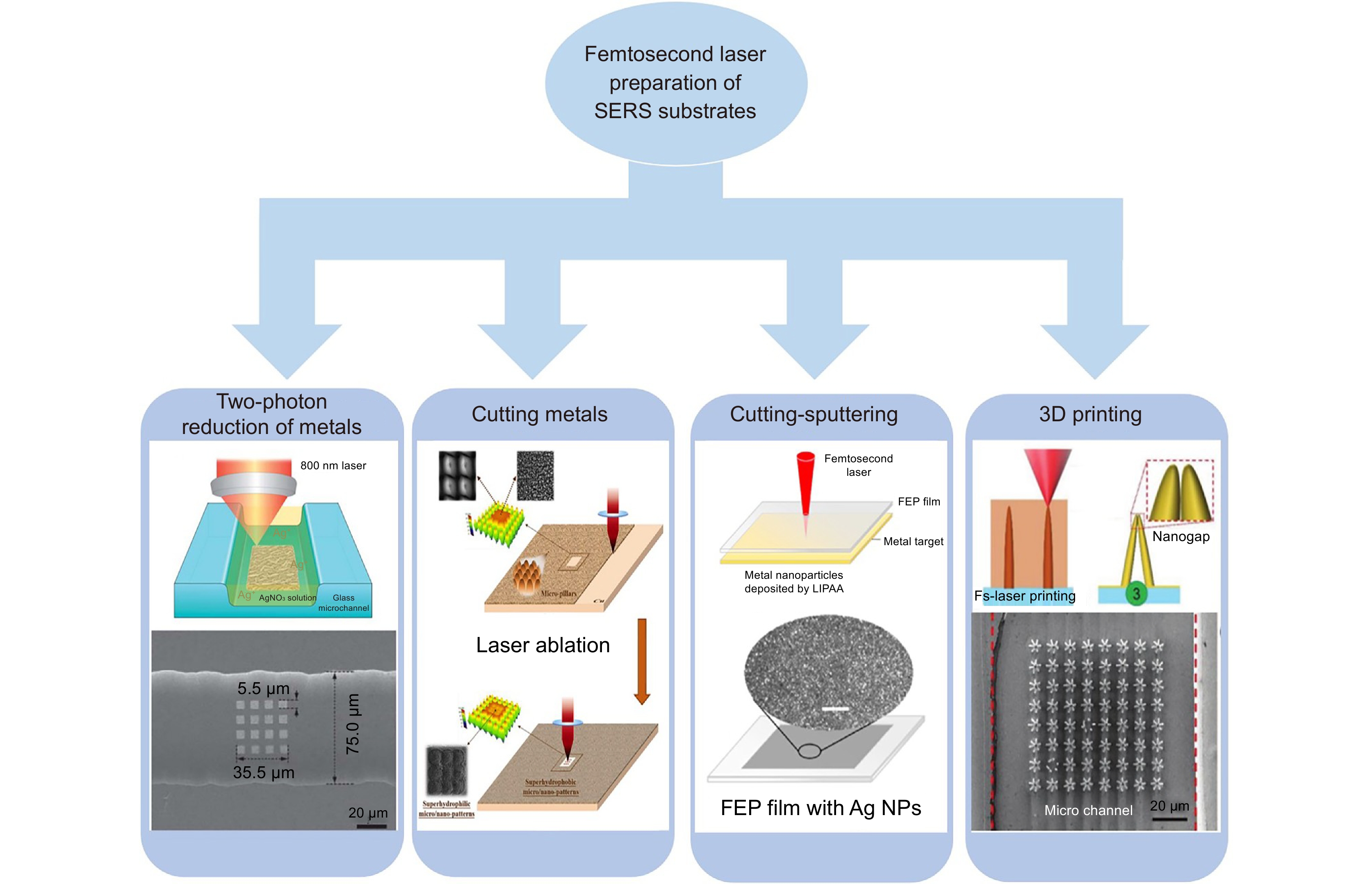
Figure 1.
Four methods of femtosecond laser preparation SERS substrate [7, 30-32]. Figure reproduced with permission from: ref. [7] © Wiley; ref. [30-31] © Elsevier; ref. [32] © The Royal Society of Chemistry
-
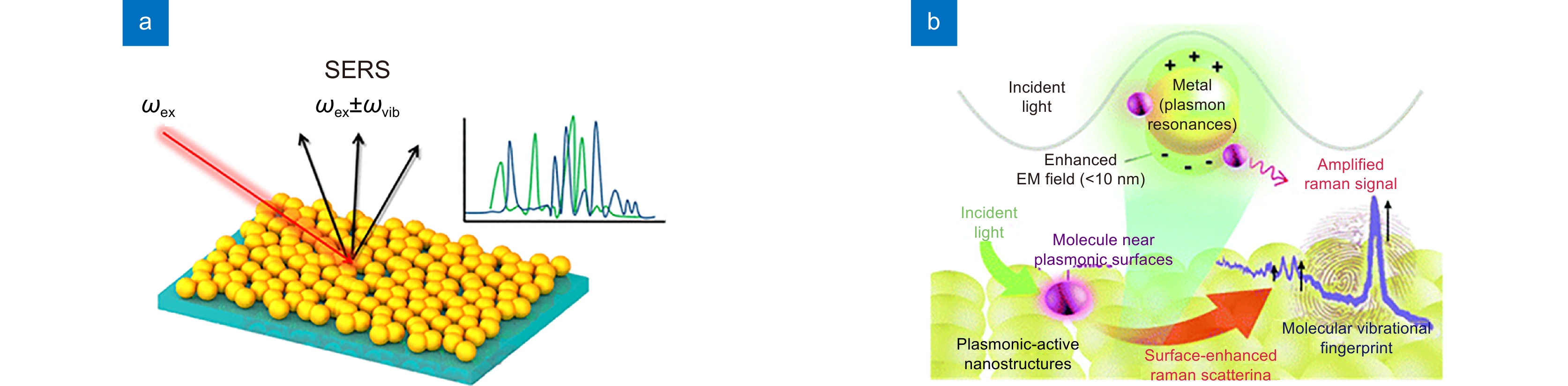
Figure 2.
SERS principle. (a) Inelastic light scattering of molecules on corrugated metal surfaces[33]; (b) localized surface plasmon resonances (LSPRs) on the surface of precious metals[36]. Figure reproduced with permission from: (a) ref. [33] © American Chemical Society; (b) ref. [36] © The Royal Society of Chemistry
-
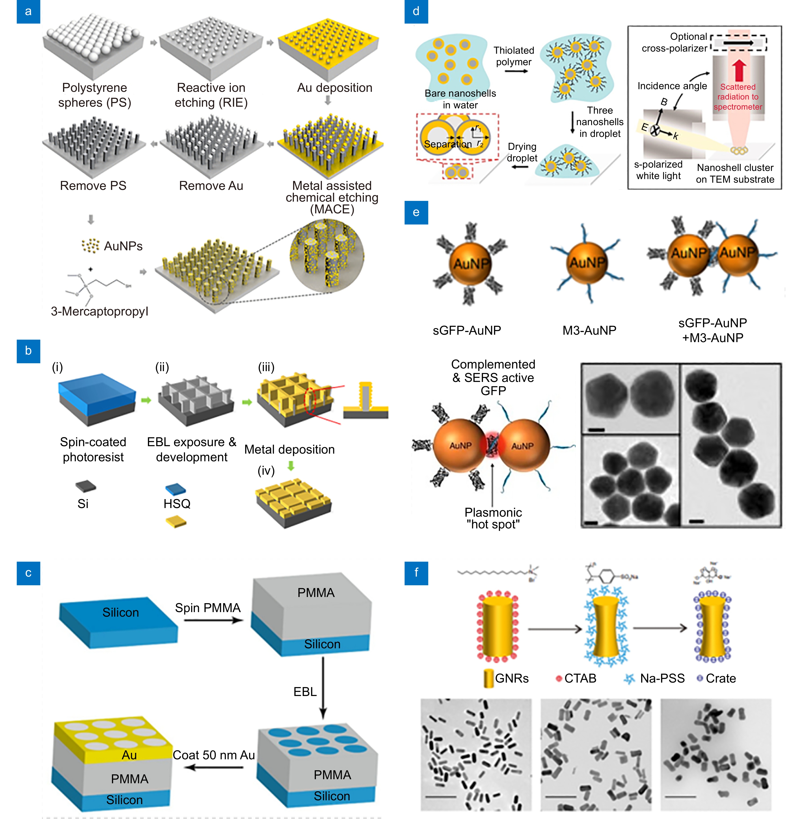
Figure 3.
Preparation of SERS microstructures by top-down micromachining and particle self-assembly. (a) RIE[45]; (b, c) EBL[46-47]; (d-f) Nanoparticle self-assembly[48-50]; Scale bar: (e) 20 nm; (f) 200 nm. Figure reproduced with permission from: (a) ref. [45] © American Chemical Society; (b) ref. [46], (e) ref. [49] and (f) ref. [50] © under a Creative Commons Attribution-NonCommercial-No- Derivatives 4.0 International License; (c) ref. [47] © American Chemical Society; (d) ref. [48] © The American Association for the Advancement of Science
-
Figure 4.
Preparation of SERS microstructure by microcolumn self-assembly methods. (a) Self-assembly of gold nanopillars[55]; (b) Self-assembly of polymer-silver micropillars[56]; (c) Self-assembly of polymer-silver micropillars[57]; (d) Self-assembly of silver micropillars[58]; (e) Self-assembly of polymer-gold micropillars[59]. Figure reproduced with permission from: (a) ref. [55] and (e) ref. [59] © American Chemical Society; (b) ref. [56], (c) ref. [57] and (d) ref. [58] © Wiley
-
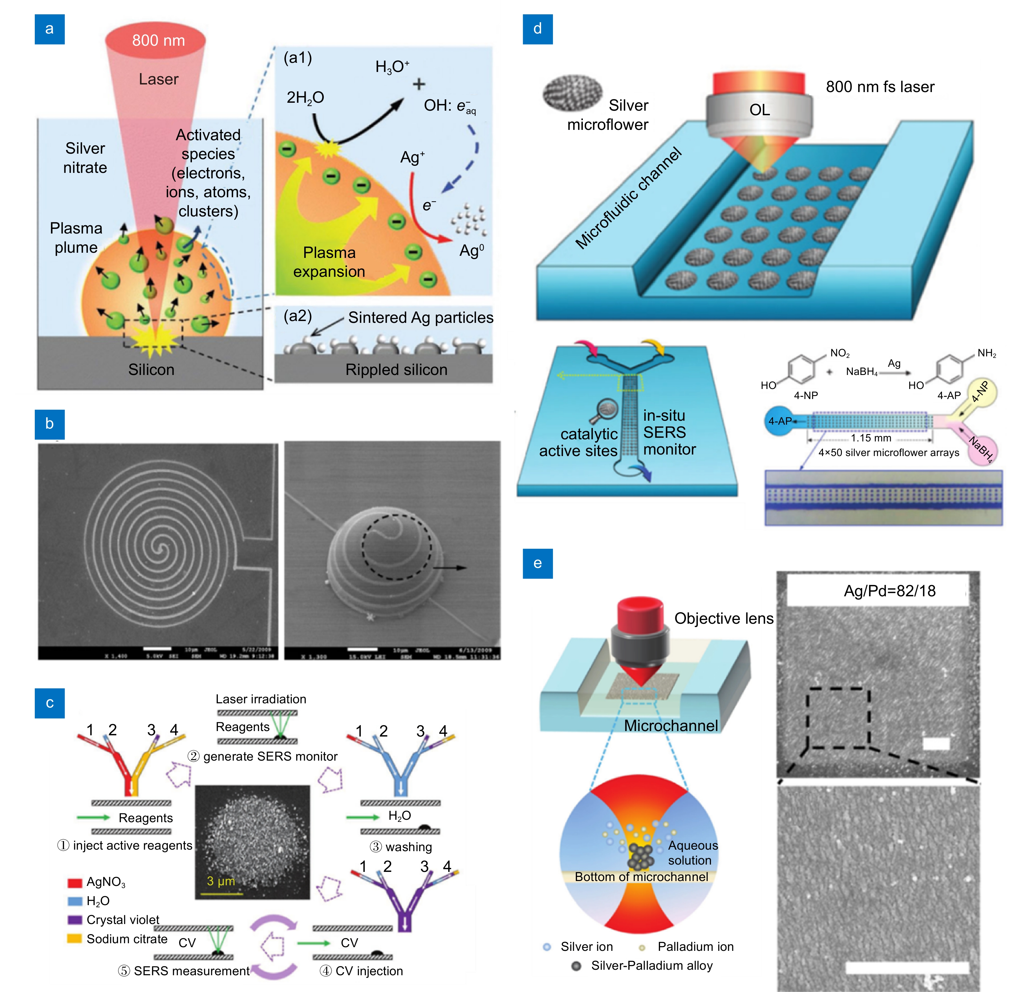
Figure 5.
Femtosecond two-photon reduction to prepare SERS substrates. (a) Two-photon reduction principle[70]; (b)Two-photon reduced silver microwire[71]; Scale bar: (b) 10 μm; (e) 1 μm. Figure reproduced with permission from: (a) ref. [70], (b) ref. [71], (c) ref. [74] and (e) ref. [71] © Wiley; (d) ref. [72] © The Royal Society of Chemistry
-
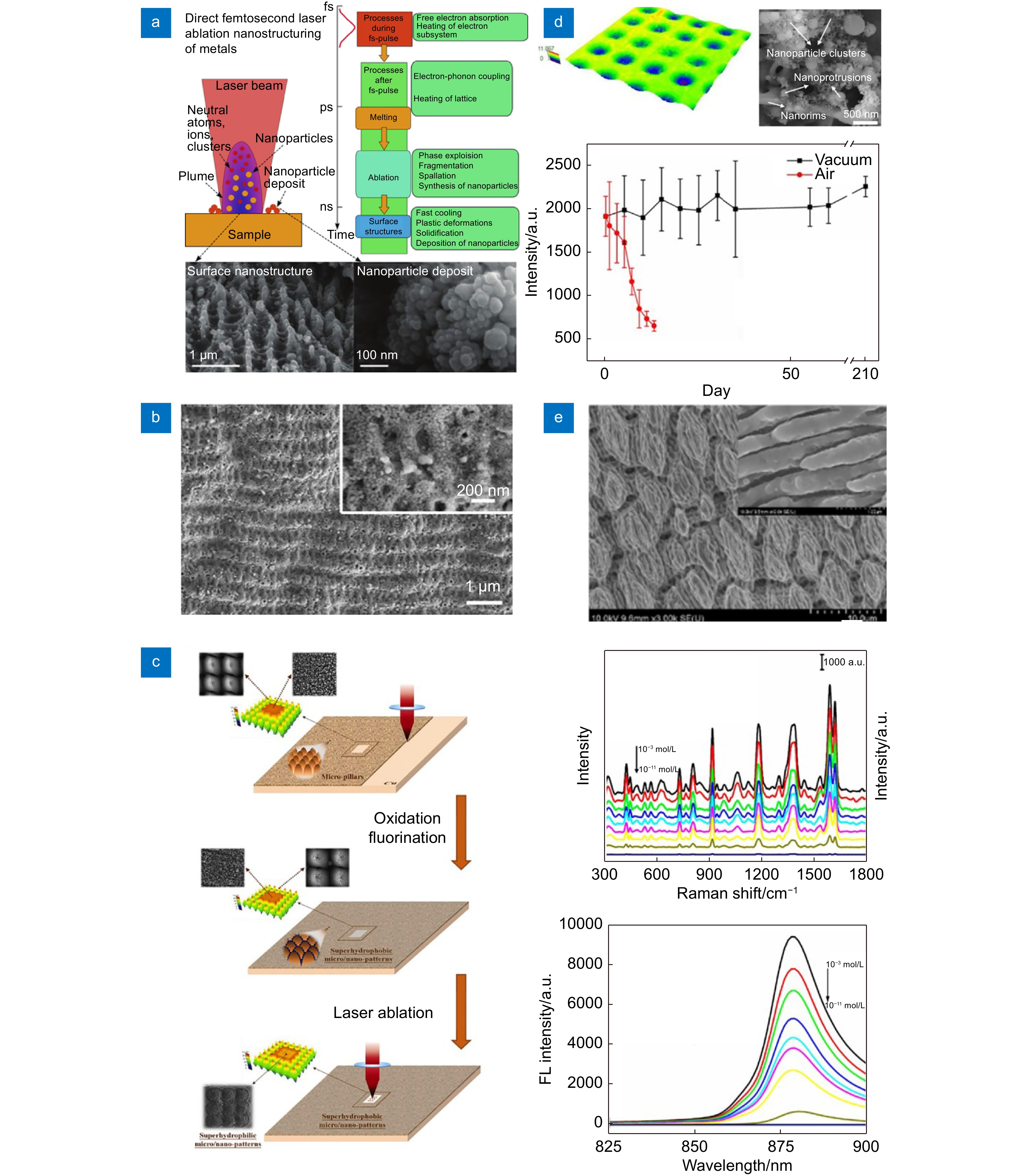
Figure 6.
Femtosecond laser cutting metal to prepare SERS substrate. (a) Femtosecond laser directly ablated metal surface forming nanostructure principle [80]; (b) Ag periodic surface[91]; (c) Superhydrophilic - superhydrophobic patterned substrate structures were prepared directly on copper surface [30]; (d) S-Ag-Ar substrate[92]; (e) Titanium alloy SERS substrate[93]. Figure reproduced with permission from: (a) ref. [80] © Elsevier; (b) ref. [91], (c) ref. [30] and (d) ref. [92] © Elsevier; (e) ref. [93] © under a Creative Commons Attribution-NonCommercial-No- Derivatives 4.0 International License
-
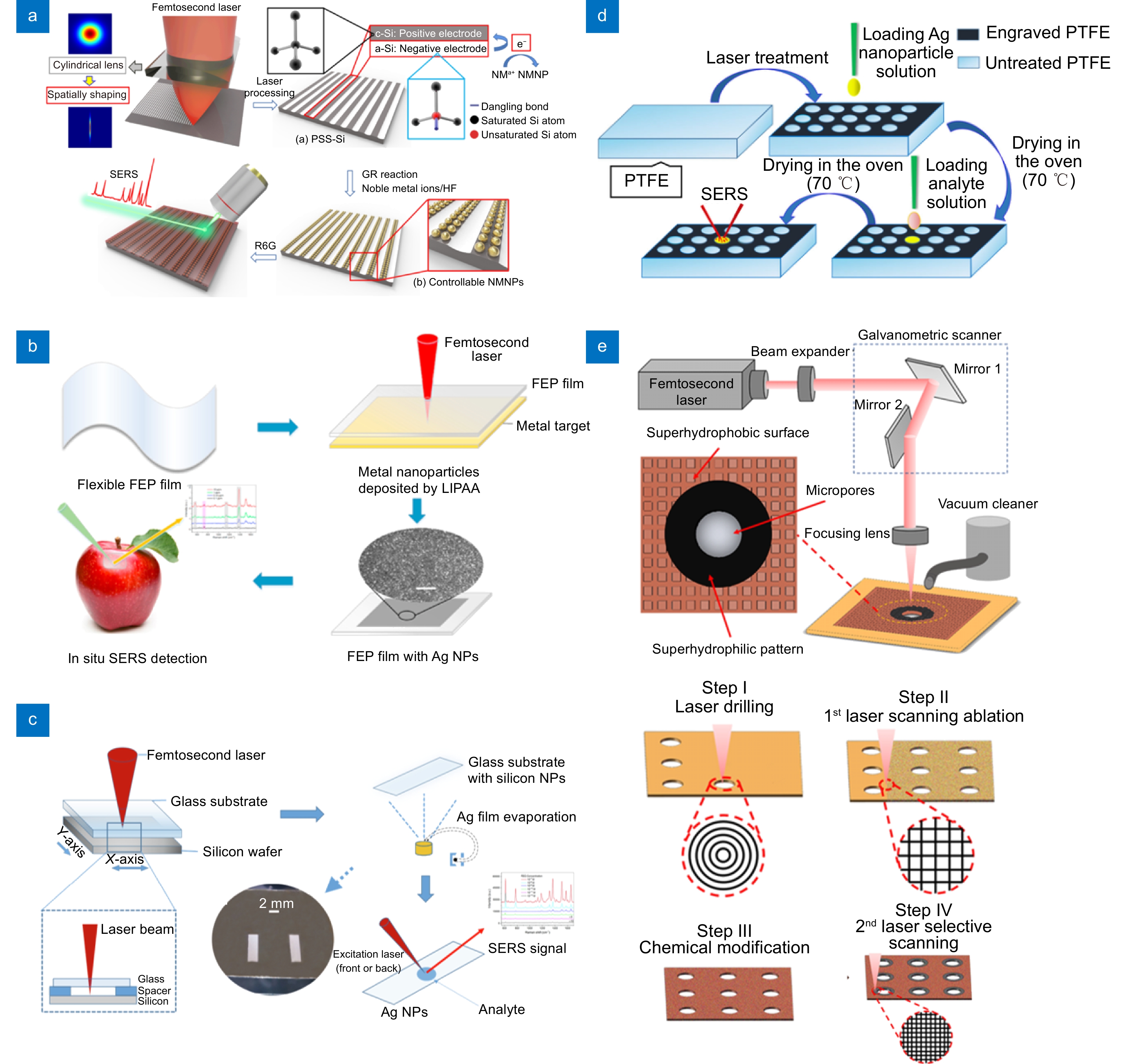
Figure 7.
Femtosecond laser cutting-sputtering to prepare a SERS substrate. (a) Large area SERS substrate[105]; (b) Flexible transparent SERS substrate[31]; (c) Glass SERS substrate[106]; (d) Hydrophobic-superhydrophobic SERS substrate[107]; (e) Superhydrophobic-hydrophilic SERS substrate[108] . Figure reproduced with permission from: (a) ref. [108], (b) ref. [31] and (c) ref. [106] © Elsevier; (d) ref. [107] © BioMed Central Ltd unless otherwise stated; (e) ref. [108] © American Chemical Society
-
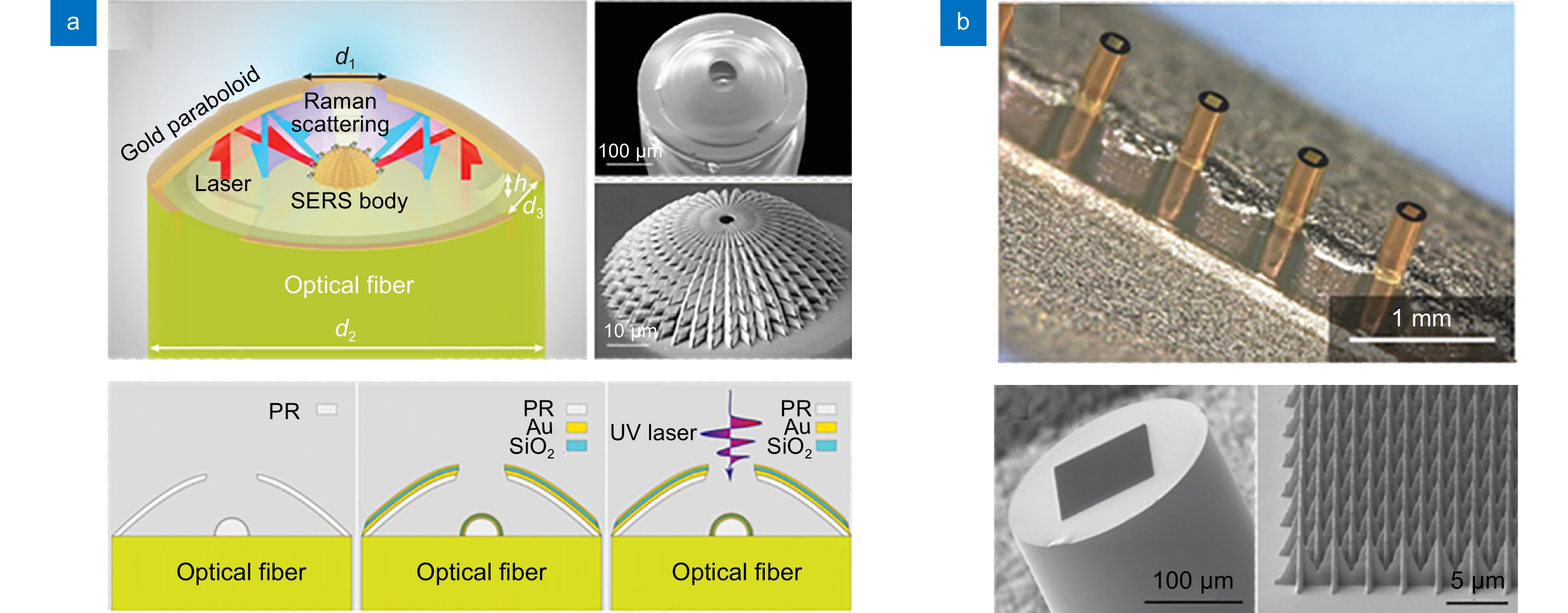
Figure 8.
Two-photon direct writing combined metal evaporation. (a, b) 3D SERS structure of fiber surface [121-122]. Figure reproduced with permission from: (a) ref. [121] © under a Creative Commons Attribution-NonCommercial-No-Derivatives 4.0 International License; (b) ref. [122] © Wiley
-
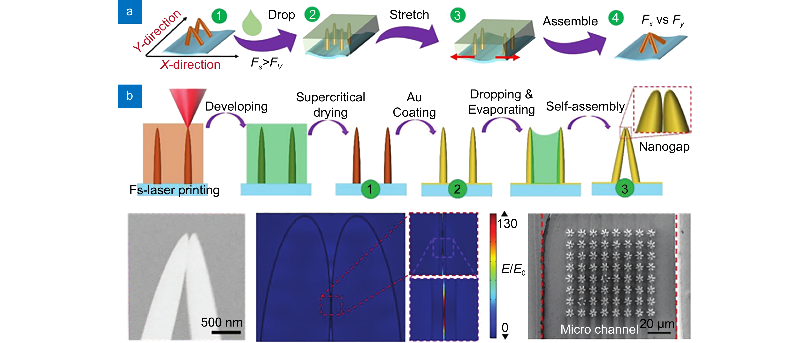
Figure 9.
Femtosecond laser processing capillary self-assembly to prepare SERS substrate. (a) Capillary force self-assembly[126]; (b) Three-dimensional SERS structure based on capillary force self-assembly microchannels[7]. Figure reproduced with permission from: (a) ref. [126] © American Chemical Society; (b) ref. [7] © Wiley


